Scars are the result of skin repair after injury. Surgery, burns, scalds, trauma, acne, infection, animal bites, and other causes may all leave scars. Different scars vary in appearance, how they develop over time, treatment difficulty, and risk of recurrence. The scar type must be identified first before appropriate care and treatment can be chosen.
Clinically, scars are often classified according to their shape and functional impact, such as hypertrophic scars, atrophic scars, contracture scars, superficial scars, depressed scars, linear scars, webbed scars, bridge scars, and others.
For most people, the most practical way to classify scars is to look at four points: whether the scar is raised, whether it is depressed, whether it causes pulling or deformity, and whether it extends beyond the original wound.
Superficial scars: mainly color change, with mild texture changes
Superficial scars usually occur after shallow skin injuries, such as minor abrasions, superficial scalds, or pigment changes after mild inflammation. Their main feature is a local color change, which may appear red, dark, or pale. The skin surface may be slightly rough or slightly shiny, but generally there is no obvious elevation or depression.
This type of scar has little effect on function and mainly affects appearance. In the early stage, sun protection is important to avoid worsening hyperpigmentation. After the wound has closed, silicone gel, moisturizing repair products, or treatments to lighten pigmentation may be used under a doctor's guidance. Most superficial scars gradually fade over time.
The treatment principle is mainly conservative care, focusing on reducing irritation, controlling inflammation, sun protection, and waiting for natural recovery. If hyperpigmentation remains obvious, energy-based light or laser treatment, chemical peeling, or topical medication may be considered, but a doctor needs to assess skin color, location, and skin sensitivity.
Linear scars: most common after surgical incisions
Linear scars are common after surgical incisions, knife cuts, and sutured wounds. Ideally, they appear as a thin line, with the color gradually approaching that of the surrounding skin and a flat, soft texture. Factors that affect the appearance of a linear scar include incision direction, suturing technique, wound tension, infection, individual constitution, and postoperative care.
If the incision is close to the direction of the skin tension lines, the scar is usually finer. If the incision is located in areas with greater tension, such as the front of the chest, shoulders and back, or near joints, the scar may widen, thicken, or even develop into a hypertrophic scar.
Early care for linear scars focuses on tension reduction and sun protection. After the wound has closed, tension-reducing tape, silicone sheets, or silicone gel can be used to reduce continuous pulling. For linear scars that have already widened, meticulous surgical revision, laser treatment, or combined treatment may be considered after the scar has stabilized.
Hypertrophic scars: raised scars confined to the original wound area
Hypertrophic scars are common pathological scars. They usually appear weeks to months after wound healing and present as redness, elevation, hardening, itching, pain, or tightness. One important feature is that they are usually confined to the original wound area.
Hypertrophic scars are common after burns, scalds, surgical incisions, trauma, and infected wounds. They also tend to occur in high-tension areas such as the front of the chest, shoulders, upper arms, and near joints. Adolescents and young adults have active tissue repair and are at higher risk.
The treatment principle is to control overgrowth early and promote scar maturation. Common methods include silicone gel, silicone sheets, pressure therapy, tension-reducing care, laser treatment, and intralesional medication injections. If the scar has stabilized but has an obvious effect on appearance or function, surgical repair may be considered. Hypertrophic scars may still overgrow again after simple excision, so postoperative tension reduction, silicone treatment, pressure therapy, or energy-based light/laser treatment is usually needed.
 View full-size image
Combined-treatment outcome
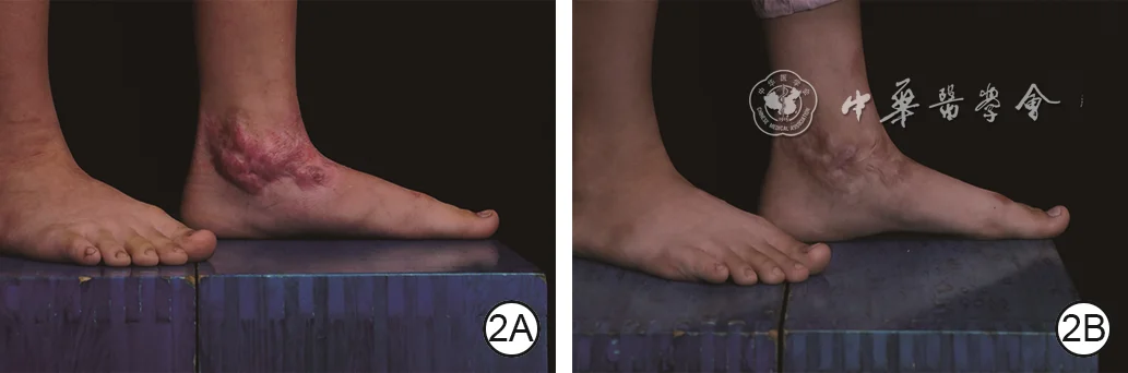
A hypertrophic scar of the left ankle was treated with pressure therapy combined with narrow-band intense pulsed light; after nine months, scar thickness and vascular congestion were reduced.
- 2ABefore treatment, the scar is markedly raised with prominent vascular congestion.
- 2BAfter nine months of treatment, scar thickness and vascular congestion have decreased.
Image source:Zeng Y, Wang C, Lin HY, et al. Clinical efficacy of pressure therapy combined with narrow-band intense pulsed light for hypertrophic scars. Chin J Burns Wounds. 2026;42(4):324–331. DOI: 10.3760/cma.j.cn501225-20251211-00518
Keloids: persistent scars that extend beyond the original wound area
Keloids are a more stubborn type of pathological scar. Their typical feature is extension beyond the original injury, spreading into surrounding normal skin, with lump-like, nodular, cord-like, or crab-claw-like growth. They are often accompanied by obvious itching, stinging pain, distending pain, and a pulling sensation, and may continue to enlarge after friction, scratching, or inflammatory irritation.
Common sites for keloids include the earlobes, lower jaw, anterior chest, shoulders and back, and upper arms. Ear piercing, acne, minor trauma, surgery, and burns can all trigger them. They are closely related to genetic predisposition, skin color, local inflammation, skin tension, and individual constitution.
The treatment principle is combined treatment and prevention of recurrence. Simple surgical excision has a high risk of recurrence because the new incision itself may again trigger scar overgrowth. Clinically, treatment is often selected according to location and size, including corticosteroid injections, surgery combined with radiotherapy, silicone therapy, laser treatment, cryotherapy, or combined drug treatment. Treatment strategies also differ for earlobe keloids and anterior chest keloids, and require individualized planning.
Atrophic scars: skin becomes thinner and lies below the surrounding skin
Atrophic scars appear as localized thinning of the skin, poor elasticity, a pale or shiny color, and a surface that lies below the surrounding skin. They are common after acne, chickenpox, trauma, infection, or long-term inflammation. Acne pits are one typical type of atrophic scar.
The formation of atrophic scars is related to loss of dermal collagen and atrophy of subcutaneous tissue. They usually do not cause obvious itching or pain, but they can make the skin surface uneven, especially on the face, where they may significantly affect appearance.
The treatment principle is to stimulate collagen regeneration and improve skin depressions and texture. Common methods include fractional laser, radiofrequency microneedling, microneedling, subcision, filler treatment, chemical peeling, and surgical excision with suturing. Different types of acne pits require different treatment approaches. Ice-pick, boxcar, and rolling acne scars are suited to different treatment plans, and multiple treatment sessions are usually needed.
Depressed scars: indentations caused by localized tissue loss
Depressed scars overlap with atrophic scars, but they place more emphasis on localized tissue loss. Trauma, infection, surgical excision, deep acne, or rupture of an abscess may all leave a depression. Their features are a localized hollow; the edges may be clearly defined or uneven.
Mild depressions can be treated with laser, radiofrequency, microneedling, and similar methods to stimulate collagen repair. Deeper depressions may require filler materials, autologous fat grafting, subcutaneous release, or surgical repair. If adhesions are present at the base of the depression, surface treatment alone has limited effect, and the deep traction must be addressed first.
 View full-size image
Clinical example
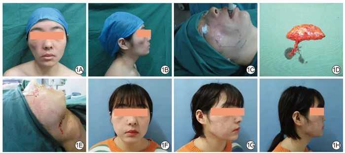
A right facial depressed scar was reconstructed with a vascularized anterolateral thigh free fat flap; facial contour and symmetry were improved at nine months.
- 1APreoperative frontal view showing depression of the right face with hypertrophic scarring on the skin surface.
- 1BPreoperative right lateral view showing soft-tissue volume deficiency and contour depression.
- 1CIntraoperative markings outline the depressed area and define the recipient-site incision position and length.
- 1DThe vascularized anterolateral thigh free fat flap immediately after harvest.
- 1EImmediate appearance after transfer of the fat flap to the facial recipient site and wound closure.
- 1FFrontal view at nine months showing flap survival and improved facial symmetry.
- 1GRight oblique view at nine months showing increased fullness of the right facial contour.
- 1HRight lateral view at nine months showing improvement of the depression and a more balanced facial contour.
Image source:Li H, Du YT, He T, et al. Clinical efficacy of anterolateral thigh free fat flap transplantation with vascular anastomosis for reconstructing facial depressed scars. Chin J Burns Wounds. 2025;41(7):665–672. DOI: 10.3760/cma.j.cn501225-20241227-00510
The treatment principle is to choose the approach according to the depth and degree of adhesion. Superficial depressions focus on stimulating regeneration, while deep depressions focus on releasing adhesions and restoring tissue volume.
Contracture scars: scars that pull on the skin and affect function
Contracture scars are common after deep burns, scalds, severe trauma, and large areas of skin loss. The main problem is scar contraction, which pulls on the surrounding skin, tendons, or joints and leads to restricted movement, deformity, or functional impairment.
Common manifestations include inability to fully straighten the fingers, limited movement of the elbow or knee, difficulty turning the neck, traction of the corner of the mouth or eyelid, and axillary scars affecting elevation of the upper limb. In children, contracture scars may also affect growth and development.
The treatment principle is to prioritize function. In the early stage, attention should be paid to pressure therapy, splinting or bracing, rehabilitation training, stretching exercises, and skin care. Once an obvious contracture has formed, surgical release is often needed, combined with repair methods such as skin grafting, flap transfer, and tissue expansion. Postoperative rehabilitation is also crucial; otherwise, the contracture may recur.
Webbed scars: traction bands forming between skin folds
Webbed scars often occur in areas such as the spaces between fingers, axilla, neck, elbow crease, popliteal fossa, and corners of the mouth. Like a web, they connect the skin on both sides and limit local unfolding and movement. They are relatively common after burns, and may also occur after trauma or surgery.
Their main problems are traction and functional limitation. For example, a webbed scar between the fingers can affect finger separation, a webbed axillary scar can affect abduction of the upper limb, and a webbed neck scar can affect raising the head and turning the neck.
The treatment principle is mainly to relieve traction and restore range of motion. Mild cases may be managed with rehabilitation training, bracing, and pressure therapy. Obvious webbed scars usually require surgical release, using methods such as Z-plasty, local flaps, or skin grafting to reconstruct the skin contour. Continuous rehabilitation and recurrence-prevention care are also needed after surgery.
Bridging scars: scars that cross over the skin surface like a bridge
Bridging scars are more common in situations involving poor wound healing, infection, irregular skin loss, or abnormal scar adhesion. They appear as scar tissue spanning the skin surface like a bridge, with a depression, groove, or irregular traction in the middle.
These scars often affect appearance and may also trap dirt, undergo repeated friction, or break down. If they are located near a joint, they may also affect movement.
The treatment principle is usually surgical revision. The doctor will choose excision, resuturing, a local flap, or another repair method according to the scar's location, length, and the condition of the surrounding skin. Postoperative care also requires tension reduction, silicone gel or sheeting, pressure therapy, and sun protection.
Pigmented scars: abnormal color is more obvious than the shape
Pigmentary scars include hyperpigmentation and hypopigmentation. Hyperpigmentation appears as darkened or brownish scarring and is common after inflammation, acne, burns or scalds, and sun exposure. Hypopigmentation appears as localized whitening and is common after deep injury, cryotherapy, or damage to the skin’s melanocytes.
The treatment principle for pigmentary scars is to stabilize inflammation first, then improve the color. Early sun protection is very important. For hyperpigmentation, topical depigmenting medications, energy-based treatments, chemical peels, and other options may be considered. Hypopigmentation is more difficult to treat; depending on the situation, phototherapy, epidermal grafting, or camouflage treatment may be considered.
General Principles of Scar Treatment
There is no single standard treatment plan for scars. Decisions need to be made comprehensively based on the type, location, size, duration, symptoms, skin color, age, constitution, and individual needs.
Early scars: focus on preventing excessive growth After the wound has closed, if redness, hardening, or itching occurs, tension reduction, silicone gel or sheeting, sun protection, and follow-up evaluation should be started as early as possible.
Raised scars: focus on controlling excessive collagen deposition Hypertrophic scars and keloids often require silicone, pressure therapy, injections, laser treatment, or combination therapy.
Depressed scars: focus on stimulating regeneration and restoring volume Acne pits and depressed scars are commonly treated with laser therapy, microneedling, radiofrequency, fillers, subcision, and other methods.
Contracture scars: focus on restoring function When joints, facial features, or limb movement are affected, surgical and rehabilitation treatment should be assessed as early as possible.
Keloid treatment should emphasize recurrence prevention Surgery usually needs to be combined with other treatments, and long-term postoperative management is very important.
References
- New Scarology, edited by Huiyuan Li, Kaihua Lu, and Shuzhong Guo, Fourth Military Medical University Press.
- Monstrey S, et al. Updated scar management practical guidelines: non-invasive and invasive measures. J Plast Reconstr Aesthet Surg. 2014;67(8):1017–1025. PubMed
- Gold MH, et al. Updated international clinical recommendations on scar management: part 2—algorithms for scar prevention and treatment. Dermatol Surg. 2014;40(8):825–831. PubMed
- Zeng Y, Wang C, Lin HY, et al. Clinical efficacy of pressure therapy combined with narrow-band intense pulsed light for hypertrophic scars. Chin J Burns Wounds. 2026;42(4):324–331. DOI: 10.3760/cma.j.cn501225-20251211-00518
- Li H, Du YT, He T, et al. Clinical efficacy of anterolateral thigh free fat flap transplantation with vascular anastomosis for reconstructing facial depressed scars. Chin J Burns Wounds. 2025;41(7):665–672. DOI: 10.3760/cma.j.cn501225-20241227-00510