Scar reconstruction refers to improving scar-related appearance abnormalities, skin defects, tissue traction, and functional impairment through surgical or combined treatment methods. In addition to the appearance of the scar, greater attention is paid to skin contour, tissue thickness, mobility, organ function, and quality of life.
Some scars are only darker in color and slightly firm, and can be improved with laser treatment, injections, silicone, or pressure therapy. Some scars have already caused skin defects, local depression, eyelid ectropion, pulling at the corner of the mouth, inability to fully straighten the fingers, or limited neck movement; these require surgical reconstruction.
The difficulty of scar reconstruction lies in the fact that skin thickness, color, tension, and functional requirements differ at every site. Therefore, treatment cannot be one-size-fits-all; it must also consider how to close the wound after excision, reduce recurrence, and restore function.
The Reconstructive Ladder
The reconstructive ladder is a commonly used treatment concept in plastic surgery. Its core principle is to start with the least traumatic and simplest repair method, then choose progressively more complex reconstruction methods according to the severity of the scar.
From lower to higher levels, it can be broadly understood as:
- Non-surgical treatment
- Direct closure or simple revision
- Local flaps or Z-plasty
- Skin grafting
- Tissue expansion
- Regional flaps or distant flaps
- Free flaps and microsurgical reconstruction
- Composite tissue reconstruction and multistage repair
The higher the level on the ladder, the more complex the technique, and the greater the surgical trauma, recovery time, and postoperative management requirements. The choice of treatment is not a matter of using the most complex option; the plastic surgeon needs to solve the specific scar problem with an appropriate method under conditions that are safe, stable, and controllable.
First Level: Non-Surgical Treatment
For mild scars, early hypertrophic scars, and scars with obvious color abnormality but acceptable shape, non-surgical treatment is usually considered first. Common methods include silicone gel, silicone sheets, pressure therapy, laser treatment, intralesional scar injections, moisturizing care, sun protection, and tension-reduction management.
Non-surgical treatment is very important in the reconstructive ladder. It can make early scars softer, flatter, and lighter in color, and it can also create better conditions for later surgery.
Second Level: Simple Excision and Fine Suturing
Simple excision with fine suturing is the most basic method of scar repair. It is suitable for linear scars, widened scars, some depressed scars, or irregular scars that are limited in extent, have sufficient skin laxity, and can be closed directly after excision.
The goal of this type of surgery is to turn an originally relatively wide, uneven scar into a thinner, flatter linear scar that follows the skin’s texture more closely. The doctor will try to design the incision along natural skin creases, tension lines, or concealed areas, while closing the wound in layers to reduce surface tension.
 View full-size image
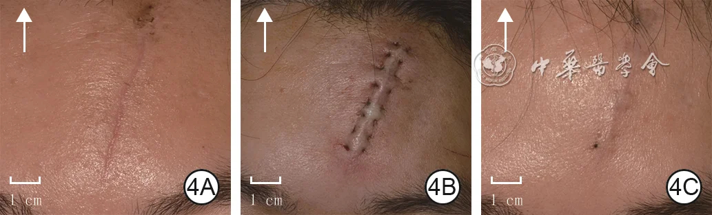
Tension reduction combined with laser treatment
A linear surgical-incision scar of the forehead was treated with horizontal buried intradermal suturing (HBIS) combined with fractional carbon dioxide laser; the scar appearance improved at one month. Arrows indicate the direction of the head.
- 4AThe forehead linear scar before combined treatment; the arrow indicates the direction of the head.
- 4BImmediate appearance after horizontal buried intradermal suturing.
- 4CScar appearance one month after combined fractional carbon dioxide laser treatment.
Image source:Lyu KY, Li YS. Mechanisms of surgical incision scar and technological innovations for scar tension reduction based on the mechano-chemo-biological theory. Chin J Burns Wounds. 2026;42(2):133–142. DOI: 10.3760/cma.j.cn501225-20251013-00424
 View full-size image
Integrated tissue remodeling
An early adult facial laceration was debrided and meticulously resutured, followed after suture removal by ion-beam tissue-remodeling treatment; two months after the second treatment, the wound scar was barely visible.
- 2AAt 3.5 days after the left facial laceration, the closure performed elsewhere appears coarse.
- 2BImmediately after debridement and meticulous resuturing, the wound is flat and well approximated.
- 2CTwo weeks after suture removal, the first ion-beam treatment is administered.
- 2DTwo months after the first ion-beam treatment, the scar is visibly improved and a second treatment is administered.
- 2ETwo months after the second ion-beam treatment, the wound scar is barely visible.
Image source:Liu W. Application of interventional tissue remodeling strategy in scar prevention and treatment. Chin J Burns Wounds. 2025;41(4):325–332. DOI: 10.3760/cma.j.cn501225-20250102-00004
 View full-size image
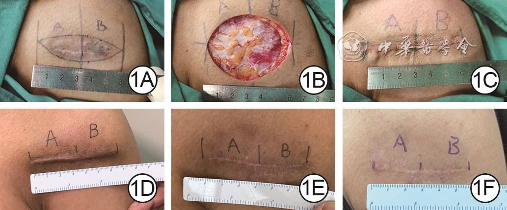
Clinical comparison
Within the same incision after fusiform excision of a right shoulder scar, healing was compared between internal sutures plus external skin sutures and internal sutures alone.
- 1AThe fusiform excision area is marked preoperatively, and the planned incision is divided into segments A and B.
- 1BThe operative field after scar excision.
- 1CAccording to random allocation, segment A receives external skin sutures after internal closure; segment B receives internal closure alone with no further treatment.
- 1DAt one month, there is no obvious difference in scar width or color between segments A and B.
- 1EAt six months, there is no obvious difference in scar width or color between segments A and B.
- 1FAt twelve months, there remains no obvious difference in scar width or color between segments A and B.
Image source:Chen ZA, Yang YT, Wang WB, et al. Prospective self-controlled clinical trial on the effects of external sutureless on skin incision healing. Chin J Burns Wounds. 2024;40(12):1143–1149. DOI: 10.3760/cma.j.cn501225-20240206-00051
Scars suitable for simple excision usually meet several conditions: the area is not large, the surrounding skin can be mobilized, removal will not cause obvious deformity, and local tension is controllable. Small facial scars, old abdominal surgical scars, and short linear scars on the limbs may all be suitable for this approach.
The limitations of simple repair are also clear. If the scar is located in a high-tension area such as the anterior chest, shoulder or back, or near a joint, it may widen again or become hypertrophic after direct closure. If a keloid is simply excised, the risk of recurrence is higher, and combined treatment with injections, radiotherapy, pressure therapy, or silicone therapy is usually needed.
 View full-size image
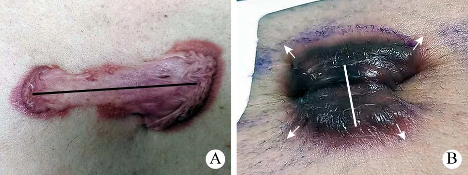
Preoperative planning
Incision planning for chest-wall keloids incorporates the lesion axis, surrounding skin folds, and local tension direction so that the incision is aligned with regional mechanical forces.
- AFor a manubrial keloid without evident surrounding skin folds, a linear incision is planned along the lesion’s long axis.
- BFor a xiphoid-region keloid, surrounding skin folds indicate the local tension direction (blue arrows); the linear incision is planned parallel to that direction.
Image source:Pan F, Wei BH, Yang W, Guo LL. Clinical outcomes of keloid core excision combined with radiotherapy in treatment of chest keloid. Academic Journal of Chinese PLA Medical School. 2022;43(5):518–523. DOI: 10.3969/j.issn.2095-5227.2022.05.005
 View full-size image
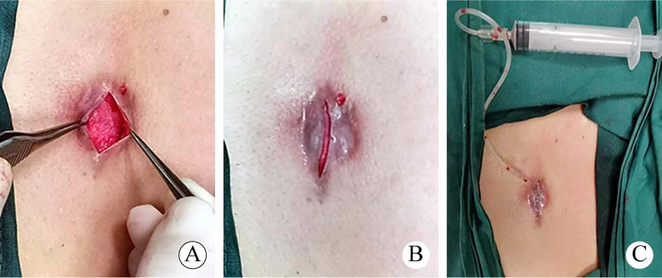
Operative example
Core excision of a chest-wall keloid with preservation of an approximately 1-mm scar flap, low-tension layered closure, and placement of a subcutaneous negative-pressure drain.
- AThe skin is incised along the long axis of the keloid; the core is dissected and removed while an approximately 1-mm scar flap is preserved.
- BImmediately after core excision, the wound edges can be approximated without obvious tension.
- CThe incision is closed in aligned layers, with a negative-pressure drain placed in the subcutaneous plane.
Image source:Pan F, Wei BH, Yang W, Guo LL. Clinical outcomes of keloid core excision combined with radiotherapy in treatment of chest keloid. Academic Journal of Chinese PLA Medical School. 2022;43(5):518–523. DOI: 10.3969/j.issn.2095-5227.2022.05.005
 View full-size image
Clinical example
Preoperative, intraoperative, and follow-up appearances of a forearm hypertrophic scar treated with conventional fusiform excision and local tension-reducing closure.
- 3ABefore revision, a linear red hypertrophic scar is visible, with puncture and suture-track marks from the initial operation on both sides.
- 3BThe fusiform incision is marked on the scar before excision.
- 3CAt one month, the healed incision is linear, mildly red, and slightly raised.
- 3DAt twelve months, the incision is flatter. It is slightly wider than at one month and remains mildly red; the earlier puncture and suture-track marks are still visible.
Image source:Ma FX, Zhang YH, Dong YC, et al. Clinical efficacy of W-plasty resection combined with remote tension-reducing suture in upper limb scar revision. Chin J Burns Wounds. 2026;42(4):350–358. DOI: 10.3760/cma.j.cn501225-20241226-00507
Third Rung: Z-Plasty and W-Plasty
Z-plasty and W-plasty are commonly used local scar revision techniques. By changing the direction of the scar, interrupting straight-line traction, and increasing local skin length, they improve linear scars, band-like scars, and mild to moderate contractures.
Z-plasty is often used for scars involving joint areas, linear traction in the neck, traction at the corner of the mouth, web-space scars of the fingers, and mild axillary contracture. It can convert a straight-line scar into a broken line while redistributing skin tension. In areas shortened by scar traction, Z-plasty can increase length in a specific direction.
W-plasty is used more often for linear facial scars, especially long straight scars. It changes a straight incision into a series of small zigzags, making the scar easier to conceal within natural skin lines and light-shadow patterns.
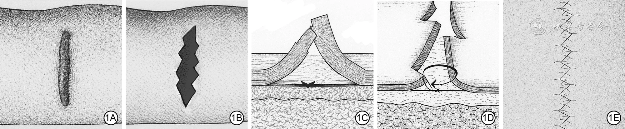 View full-size image
Technique illustration
Stepwise illustration of W-plasty excision combined with remote tension-reducing sutures.
- 1APreoperative scar.
- 1BThe wound after excision of the scar using a W-shaped design.
- 1CRemote deep dermal tension-reducing sutures.
- 1DSubcutaneous tissue closure; the arrow indicates the suture path.
- 1EFine epidermal closure.
Image source:Ma FX, Zhang YH, Dong YC, et al. Clinical efficacy of W-plasty resection combined with remote tension-reducing suture in upper limb scar revision. Chin J Burns Wounds. 2026;42(4):350–358. DOI: 10.3760/cma.j.cn501225-20241226-00507
These procedures require a high level of design precision. Angles, lengths, flap blood supply, and the direction of tension all need to be calculated. When the design is appropriate, local contour and mobility can improve significantly; when the design is poor, a new conspicuous scar or local skin necrosis may occur.
 View full-size image
Clinical example
Preoperative, intraoperative, and follow-up appearances of a linear right forearm scar treated with W-plasty excision and remote tension-reducing sutures.
- 2ABefore revision, the linear scar is slightly depressed and uneven in color, with puncture and suture-track marks from the initial operation on both sides.
- 2BThe W-shaped incision is marked on the scar before excision.
- 2CAt one month, the healed incision is linear, red, and prominently raised.
- 2DAt twelve months, the incision is markedly flatter and its color approaches the surrounding skin; no residual puncture or suture-track marks are visible.
Image source:Ma FX, Zhang YH, Dong YC, et al. Clinical efficacy of W-plasty resection combined with remote tension-reducing suture in upper limb scar revision. Chin J Burns Wounds. 2026;42(4):350–358. DOI: 10.3760/cma.j.cn501225-20241226-00507
Fourth Rung: Local Flap Repair
A local flap uses healthy skin and subcutaneous tissue near the scar, which is rotated, advanced, or transposed into the defect left after scar excision. Its advantage is that the color, texture, and thickness are close to those of the original site, so the appearance after repair is usually more natural than with skin grafting.
Local flaps are suitable for areas with high aesthetic and functional requirements, such as the face, neck, hands, and regions near joints. For example, for small to medium defects of the nasal ala, eyelids, perioral region, mandible, and periauricular area, local flaps are often considered first. For scar contractures near joints, local flaps can also provide softer, more elastic coverage tissue.
Common local flaps include advancement flaps, rotation flaps, transposition flaps, rhomboid flaps, and V-Y advancement flaps. The doctor will design the repair comprehensively according to factors such as defect size, direction of blood supply, and skin laxity.
The limitation of local flaps is that available tissue is limited. If the scar area is large, the surrounding skin is also damaged, or local skin elasticity is insufficient, a local flap alone may not be enough, and a higher rung may be needed.
Fifth Rung: Skin Grafting
Skin grafting means taking a layer of skin from another part of the body and transplanting it onto the wound surface after scar excision or contracture release. It is often used for post-burn scars, large skin defects, large wounds after contracture release, and situations where local flaps cannot provide enough coverage.
Skin grafts can be divided into split-thickness grafts and full-thickness grafts. Split-thickness grafts are easier to take and are suitable for larger wounds, but later they may show color mismatch, contraction, and texture differences. Full-thickness grafts have better texture and relatively less contraction, and are often used for small defects in delicate areas such as the face and hands, but donor-site skin is limited.
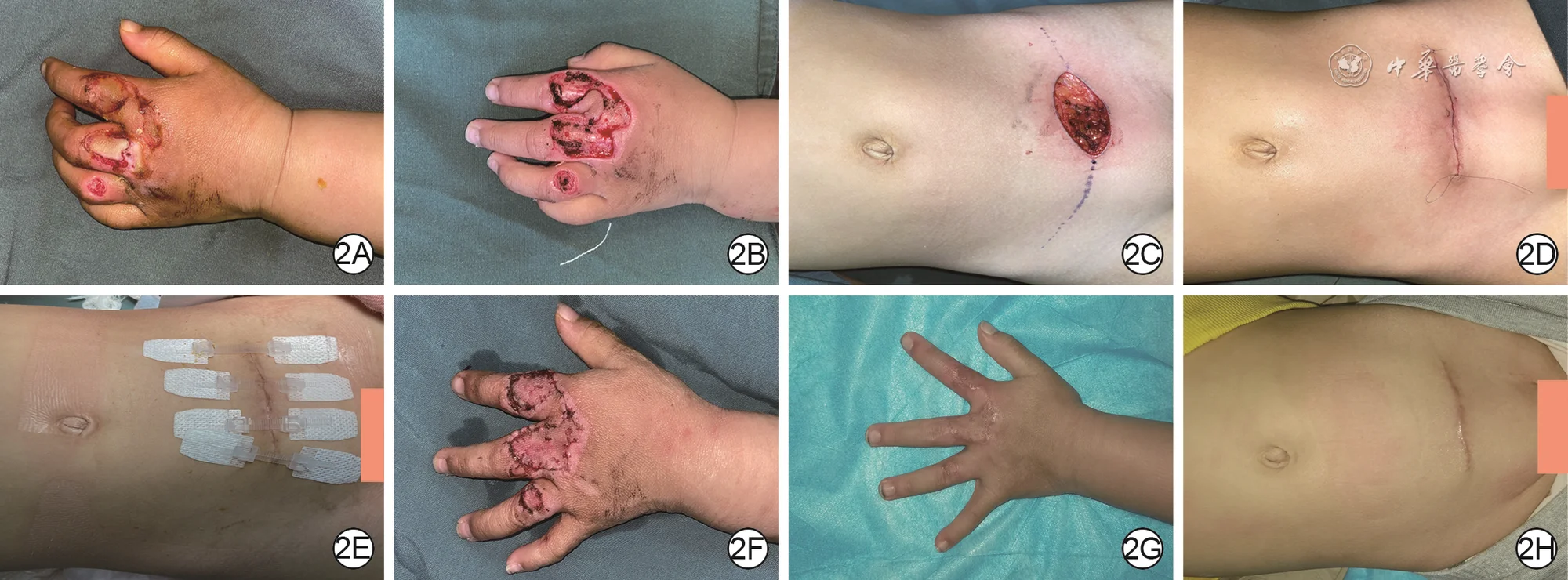 View full-size image
Clinical example
Surgical course and follow-up of a treadmill-related contact burn on a child’s left dorsal hand repaired with a full-thickness skin graft from the lower abdomen.
- 2APreoperative wounds on the dorsal index, middle, and ring fingers of the left hand.
- 2BThe left-hand wound immediately after debridement.
- 2CThe secondary lower-abdominal donor-site wound after harvesting the full-thickness skin graft.
- 2DThe donor site immediately after intradermal closure.
- 2EAt the postoperative day-7 dressing change, the donor-site incision had healed well; sutures were removed and a medical skin tension-reduction closure device was applied.
- 2FAt the postoperative day-10 dressing change, the graft on the left hand had survived.
- 2GAt six months, the graft color approximated the surrounding normal skin, with no scar contracture and a favorable hand appearance.
- 2HAt six months, the donor-site scar remained linear without obvious widening compared with the immediate postoperative appearance.
Image source:Tong L, Zhang WF, Hu XL, et al. A prospective randomized controlled study on the repair of skin and soft tissue defect in functional areas of children with full-thickness skin grafts from different sites of abdomen. Chin J Burns Wounds. 2022;38(8):744–752. DOI: 10.3760/cma.j.cn501120-20210709-00243
The advantages of skin grafting are that it can cover a large area and the technique is relatively well established. Its shortcomings are also clear: the transplanted skin lacks complete skin appendage structures, so its color and texture may differ; near joints, it may contract again; after surgery, pressure therapy, splints, and rehabilitation training are needed.
Skin grafting is commonly used for release of post-burn scar contractures, but if the functional demands of the area are high, or if tissue that is more wear-resistant and more elastic is needed, the doctor may choose flap repair.
Sixth Rung: Tissue Expansion Repair
Tissue expansion is a very important method in scar reconstruction. Its principle is to place an expander near or adjacent to the scar and inject saline regularly, allowing normal skin to be gradually expanded. After several weeks or months, the doctor then uses the expanded skin to repair the scarred area.
The greatest advantage of tissue expansion is that it repairs an adjacent area with similar skin. Tissue expansion may be used for scalp scars, facial and neck scars, trunk scars, and post-burn scars in children. The expanded skin is close in color, texture, and thickness to the surrounding area, making it especially suitable for areas where appearance is a high priority.
For example, with a scalp scar accompanied by hair loss, if skin grafting is performed directly, the repaired area will have no hair, creating an obvious difference in appearance. Through scalp expansion, hair-bearing scalp can be used to cover the scarred area, producing a more natural appearance. Facial and neck scars can also achieve better color and texture matching through expansion.
The disadvantages of tissue expansion are that the treatment period is long, repeated follow-up visits are needed for saline filling, and the local area will bulge during expansion, which may affect appearance and daily life. Complications include infection, expander exposure, skin breakdown, pain, and inadequate expansion. Patient cooperation has a major impact on the result.
Seventh rung: regional flaps and distant flaps
When local tissue is insufficient and a skin graft cannot meet functional and aesthetic requirements, regional flaps or distant flaps may be considered. A regional flap transfers tissue with its own blood supply from a nearby area; a distant flap may come from a site farther away.
These types of flaps are often used for scar defects that are large, deep, or expose important structures. For example, after scar excision, tendons, bone, joints, blood vessels, or nerves may be exposed. Simple skin grafting is unlikely to survive in such cases, so a flap with its own blood supply is needed for coverage.
Compared with a skin graft, a flap is thicker, has a better blood supply, and has stronger resistance to infection and wear. For functional areas such as the hand, foot and ankle, knee, and elbow, a flap can provide more stable soft-tissue coverage and reduce recurrent breakdown.
The trade-off with regional flaps is increased surgical complexity, and the donor site will also be left with a scar. The doctor needs to balance the reconstructive result against donor-site injury.
Eighth rung: microsurgical free flaps
Microsurgical reconstruction is an advanced stage on the reconstructive ladder. A free flap requires taking skin, fascia, muscle, or composite tissue with blood vessels from one part of the body and, under a microscope, connecting those vessels to vessels in the recipient area so that the transplanted tissue regains its blood supply.
 View full-size image
Contracture release and functional reconstruction
Release of a post-electrical-burn scar contracture of the left palm exposed vessels and tendons; coverage was achieved with a left medial plantar free flap, which maintained a favorable contour at ten months.
- 2ABefore surgery, scar contracture is present on the left palm after an electrical burn.
- 2BAfter scar excision and release, vessels and tendons are exposed in the palmar wound.
- 2CImmediate appearance after coverage of the released wound with a medial plantar free flap.
- 2DAt ten months, the flap on the left palm has a favorable contour.
Image source:Han F, Yang XK, He T, et al. Curative effects of medial plantar free flap in reconstructing electric burn wound and scar contracture in the palm. Chin J Burns Wounds. 2023;39(9):820–825. DOI: 10.3760/cma.j.cn501225-20230601-00197
 View full-size image
Microsurgical reconstruction
A left medial plantar free flap was used to reconstruct an electrical burn wound of the left palm; at six months, the palmar flap had a favorable contour and the plantar donor site showed no evident scar contracture.
- 1ABefore surgery, the left palmar electrical burn wound contains leathery necrotic tissue.
- 1BThe flap harvest area is designed on the left medial plantar region according to the wound size after debridement.
- 1CAt six months, the flap on the left palm has a favorable contour.
- 1DAt six months, no evident scar contracture is present at the left plantar donor site.
Image source:Han F, Yang XK, He T, et al. Curative effects of medial plantar free flap in reconstructing electric burn wound and scar contracture in the palm. Chin J Burns Wounds. 2023;39(9):820–825. DOI: 10.3760/cma.j.cn501225-20230601-00197
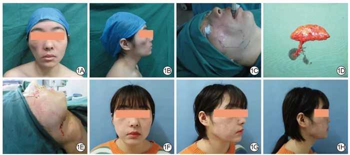 View full-size image
Clinical example
A right facial depressed scar was reconstructed with a vascularized anterolateral thigh free fat flap; facial contour and symmetry were improved at nine months.
- 1APreoperative frontal view showing depression of the right face with hypertrophic scarring on the skin surface.
- 1BPreoperative right lateral view showing soft-tissue volume deficiency and contour depression.
- 1CIntraoperative markings outline the depressed area and define the recipient-site incision position and length.
- 1DThe vascularized anterolateral thigh free fat flap immediately after harvest.
- 1EImmediate appearance after transfer of the fat flap to the facial recipient site and wound closure.
- 1FFrontal view at nine months showing flap survival and improved facial symmetry.
- 1GRight oblique view at nine months showing increased fullness of the right facial contour.
- 1HRight lateral view at nine months showing improvement of the depression and a more balanced facial contour.
Image source:Li H, Du YT, He T, et al. Clinical efficacy of anterolateral thigh free fat flap transplantation with vascular anastomosis for reconstructing facial depressed scars. Chin J Burns Wounds. 2025;41(7):665–672. DOI: 10.3760/cma.j.cn501225-20241227-00510
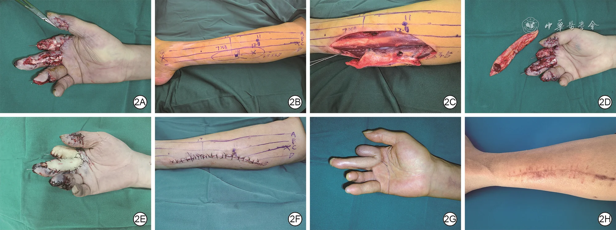 View full-size image
Clinical example
Operative sequence and nine-month follow-up of right-hand wound reconstruction using a peroneal artery perforator flap designed and harvested with the “three-source and four-line method.”
- 2AWounds of the right index and middle fingers after debridement.
- 2BThe flap is designed on the left lower leg using the “three-source and four-line method.”
- 2CTwo perforators are identified intraoperatively, both arising from the peroneal artery.
- 2DThe harvested flap is ready for transfer to the recipient site.
- 2EImmediate appearance of the recipient site after flap reconstruction.
- 2FThe left lower-leg donor site after direct approximation and closure.
- 2GAt nine months, the flap has favorable color and no obvious bulkiness.
- 2HAt nine months, a linear scar remains at the donor site.
Image source:Wu CL, Zhang Y, Cheng JN, et al. Clinical application effects of the “three-source and four-line method” in the design and harvest of anterolateral lower leg perforator flaps. Chin J Burns Wounds. 2026;42(3):252–260. DOI: 10.3760/cma.j.cn501225-20250328-00148
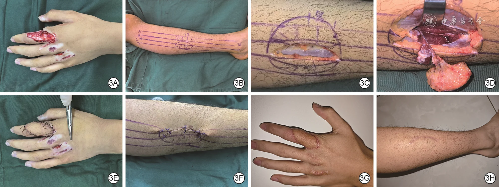 View full-size image
Clinical example
Operative sequence and ten-month follow-up of left-hand wound reconstruction using the “three-source and four-line method” to identify an alternative perforator and adjust a superficial peroneal artery perforator flap.
- 3AWound of the left index finger after debridement.
- 3BThe flap is designed on the right lower leg using the “three-source and four-line method.”
- 3CAfter an alternative perforator is detected, the flap harvest position is adjusted.
- 3DIntraoperative tracing shows that the alternative perforator arises from the superficial peroneal artery.
- 3EImmediate appearance of the recipient site after flap reconstruction.
- 3FThe right lower-leg donor site after direct approximation and closure.
- 3GAt ten months, the flap has favorable color and no obvious bulkiness.
- 3HAt ten months, a linear scar remains at the donor site.
Image source:Wu CL, Zhang Y, Cheng JN, et al. Clinical application effects of the “three-source and four-line method” in the design and harvest of anterolateral lower leg perforator flaps. Chin J Burns Wounds. 2026;42(3):252–260. DOI: 10.3760/cma.j.cn501225-20250328-00148
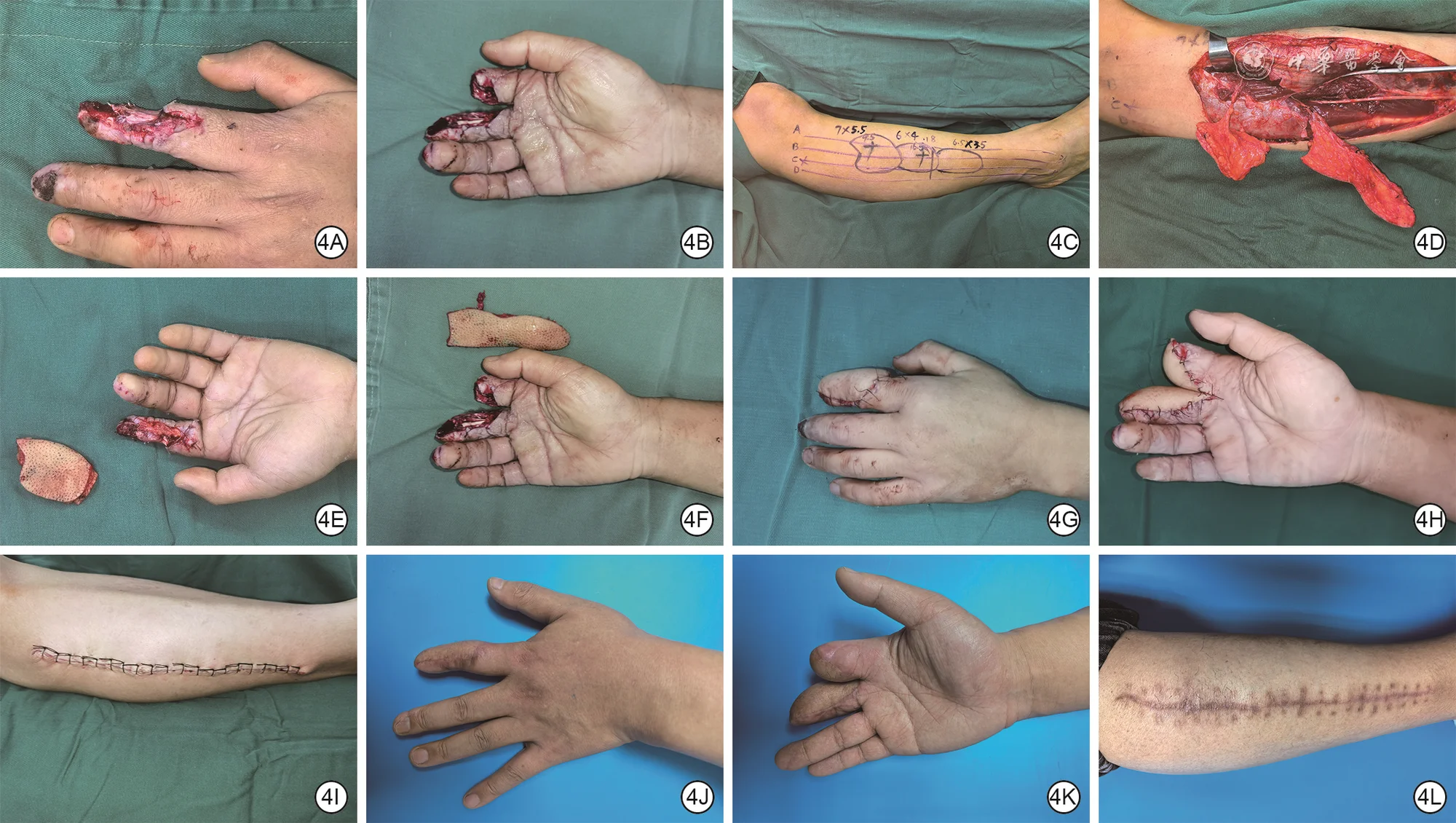 View full-size image
Clinical example
Operative sequence and eight-month follow-up of bilateral hand wound reconstruction using anterior tibial and superficial peroneal artery perforator flaps designed with the “three-source and four-line method.”
- 4ALeft index-finger wound after debridement.
- 4BRight index- and middle-finger wounds after debridement.
- 4CThe flaps are designed on the right lower leg using the “three-source and four-line method.”
- 4DTwo perforators are identified intraoperatively, arising from the anterior tibial and superficial peroneal arteries.
- 4EThe harvested flap is prepared for transfer to the left-hand recipient site.
- 4FThe harvested flap is prepared for transfer to the right-hand recipient site.
- 4GImmediate appearance of the left-hand recipient site after flap reconstruction.
- 4HImmediate appearance of the right-hand recipient site after flap reconstruction.
- 4IThe right lower-leg donor site after direct approximation and closure.
- 4JAt eight months, the left-hand flap has favorable color and no obvious bulkiness.
- 4KAt eight months, the right-hand flap has favorable color and no obvious bulkiness.
- 4LAt eight months, a linear scar remains at the donor site.
Image source:Wu CL, Zhang Y, Cheng JN, et al. Clinical application effects of the “three-source and four-line method” in the design and harvest of anterolateral lower leg perforator flaps. Chin J Burns Wounds. 2026;42(3):252–260. DOI: 10.3760/cma.j.cn501225-20250328-00148
This method is suitable for complex, large-area scar reconstruction with obvious deep tissue defects. Examples include severe post-burn soft-tissue defects of the neck, face, hands, or lower limbs; exposure of bone, tendon, or joints after scar excision; repeated surgical failures; or very poor local tissue conditions.
Microsurgery can provide, in a single procedure, a relatively large area of tissue with reliable blood supply and appropriate thickness. Depending on the need, options may include an anterolateral thigh flap, latissimus dorsi flap, anterolateral thigh perforator flap, or deep inferior epigastric artery perforator flap. Some complex reconstructions may also involve reconstruction of nerves, tendons, or bone.
Microsurgery places high demands on hospital resources, surgeon experience, and postoperative monitoring. After surgery, the flap’s blood supply must be observed closely to prevent vascular crisis. The recovery period is relatively long, but for functional reconstruction of complex scars, it is often a very valuable option.
Ninth rung: composite tissue and multi-stage reconstruction
Some scars cause problems beyond the skin surface. For example, after a severe facial burn, there may be skin loss, eyelid ectropion, perioral contracture, nasal ala deformity, hair loss, and psychological stress all at the same time. Severe hand scars may be combined with tendon adhesions, joint stiffness, nerve injury, and soft-tissue defects.
This situation usually requires multi-stage reconstruction. The first stage may focus on releasing contracture and restoring basic function; the second stage on improving tissue coverage; the third stage on refining appearance; and later stages may include laser treatment, fat grafting, hair transplantation, texture improvement, or secondary fine revision.
Composite tissue reconstruction emphasizes overall planning. The doctor will assess facial proportions, organ function, joint mobility, donor-site conditions, the patient’s age, occupational needs, psychological state, and more. In addition, treatment of complex scars takes a long time, so patients need reasonable expectations.
Scar Reconstruction Q&A
How do you choose the appropriate rung of the reconstructive ladder?
The choice of repair method mainly depends on several factors:
Scar size and depth Small linear scars may be suitable for simple excision, while large burn scars may require skin grafting, tissue expansion, or flap reconstruction.
Whether function is affected Scars that affect movement of the fingers, neck, armpit, eyelids, corners of the mouth, or joints have a higher priority for functional recovery.
Condition of the surrounding skin If the surrounding skin is lax and healthy, a local flap or direct closure may be chosen; if the surrounding skin is also scarred, a higher rung of the reconstructive ladder may be needed.
Whether recurrence is likely Keloids, scars on the anterior chest, and scars on the shoulders or back have a higher risk of recurrence after surgery and often require combined treatment.
Aesthetic requirements and site-specific features The face, neck, and hands require greater attention to color, thickness, texture, and fine function.
Whether the patient can cooperate with long-term management Tissue expansion, pressure therapy, splint-based rehabilitation, and postoperative care after microsurgery all require a high degree of cooperation.
Overall, the choice of treatment method is the result of comprehensive consideration and is not determined by the patient alone or by the doctor alone.
Why does scar reconstruction require postoperative management?
After scar reconstruction is completed, the new incision will still go through the process of scar formation, and postoperative management directly affects the final outcome.
Common postoperative scar management includes: tension reduction, silicone therapy, pressure therapy, sun protection, laser treatment, injections, massage, splints, rehabilitation exercises, and regular follow-up visits. Scars near joints especially require movement training to prevent contracture from recurring. Patients with keloids need longer-term observation and, when necessary, combined radiotherapy, injections, or pressure therapy to reduce the risk of recurrence.
Reconstructive surgery addresses structural problems, while long-term care helps determine whether the scar remains stable. Neglecting postoperative management can lead to unwanted widening, hypertrophy, pigmentation, contracture, or recurrence, even when the operation was well planned.
References
- New Scarology, edited by Huiyuan Li, Kaihua Lu, and Shuzhong Guo, Fourth Military Medical University Press.
- Monstrey S, et al. Updated scar management practical guidelines: non-invasive and invasive measures. J Plast Reconstr Aesthet Surg. 2014;67(8):1017–1025. PubMed
- Gold MH, et al. Updated international clinical recommendations on scar management: part 2—algorithms for scar prevention and treatment. Dermatol Surg. 2014;40(8):825–831. PubMed
- Lyu KY, Li YS. Mechanisms of surgical incision scar and technological innovations for scar tension reduction based on the mechano-chemo-biological theory. Chin J Burns Wounds. 2026;42(2):133–142. DOI: 10.3760/cma.j.cn501225-20251013-00424
- Han F, Yang XK, He T, et al. Curative effects of medial plantar free flap in reconstructing electric burn wound and scar contracture in the palm. Chin J Burns Wounds. 2023;39(9):820–825. DOI: 10.3760/cma.j.cn501225-20230601-00197
- Liu W. Application of interventional tissue remodeling strategy in scar prevention and treatment. Chin J Burns Wounds. 2025;41(4):325–332. DOI: 10.3760/cma.j.cn501225-20250102-00004
- Pan F, Wei BH, Yang W, Guo LL. Clinical outcomes of keloid core excision combined with radiotherapy in treatment of chest keloid. Academic Journal of Chinese PLA Medical School. 2022;43(5):518–523. DOI: 10.3969/j.issn.2095-5227.2022.05.005
- Li H, Du YT, He T, et al. Clinical efficacy of anterolateral thigh free fat flap transplantation with vascular anastomosis for reconstructing facial depressed scars. Chin J Burns Wounds. 2025;41(7):665–672. DOI: 10.3760/cma.j.cn501225-20241227-00510
- Ma FX, Zhang YH, Dong YC, et al. Clinical efficacy of W-plasty resection combined with remote tension-reducing suture in upper limb scar revision. Chin J Burns Wounds. 2026;42(4):350–358. DOI: 10.3760/cma.j.cn501225-20241226-00507
- Tong L, Zhang WF, Hu XL, et al. A prospective randomized controlled study on the repair of skin and soft tissue defect in functional areas of children with full-thickness skin grafts from different sites of abdomen. Chin J Burns Wounds. 2022;38(8):744–752. DOI: 10.3760/cma.j.cn501120-20210709-00243
- Wu CL, Zhang Y, Cheng JN, et al. Clinical application effects of the “three-source and four-line method” in the design and harvest of anterolateral lower leg perforator flaps. Chin J Burns Wounds. 2026;42(3):252–260. DOI: 10.3760/cma.j.cn501225-20250328-00148
- Chen ZA, Yang YT, Wang WB, et al. Prospective self-controlled clinical trial on the effects of external sutureless on skin incision healing. Chin J Burns Wounds. 2024;40(12):1143–1149. DOI: 10.3760/cma.j.cn501225-20240206-00051