After a wound has healed, redness, elevation, and hardening are medically often classified as pathological scarring, which mainly includes hypertrophic scars and keloids.
Both are related to excessive skin repair and excessive collagen deposition, and both can affect appearance and even cause itching, pain, or a pulling sensation.
Hypertrophic scars are usually confined to the original wound area and may gradually become flatter and softer over time. Keloids are more likely to extend beyond the original area of injury into the surrounding normal skin, and they also carry a higher risk of recurrence.
During diagnosis or treatment, confusing the two may affect the choice of treatment plan and may even increase the risk of recurrence.
Hypertrophic Scars
A hypertrophic scar is a scar formed by an overly strong local repair response during wound healing. Common features include a raised, red, hardened scar; some people also have itching, stinging pain, a burning sensation, or a feeling of tightness.
Hypertrophic scars usually appear within the area of the original wound. For example, if a surgical incision is a line, the hypertrophic scar usually thickens along that incision; if a burn or scald wound is located in a certain area, the hypertrophic scar is also mainly concentrated in the original wound area.
Hypertrophic scars often occur after surgery, burns, scalds, trauma, or infected wounds, and they are especially likely to appear in areas with greater skin tension, such as the front of the chest, shoulders, upper arms, and near joints. Adolescents and young adults have strong tissue repair capacity and active fibroblasts, so their likelihood of developing hypertrophic scars is also relatively higher.
Hypertrophic scars often become gradually noticeable several weeks to several months after a wound has healed. In the early stage, they tend to be red, firm in texture, and obviously raised. Over time, some hypertrophic scars enter a maturation phase, with the color fading, the texture softening, the thickness decreasing, and itching and pain easing. This process may last six months to more than one year.
Keloids
A keloid is a more stubborn type of pathological scar with benign tumor-like growth characteristics. Its typical feature is that it extends beyond the original wound area into the surrounding normal skin and shows persistent, mass-like growth.
A keloid may start as a small hard nodule and gradually become larger and thicker, with its borders expanding outward. Its shape may be round, oval, nodular, or plaque-like, and it may also have a crab-claw-like appearance. The surface is often shiny, the texture tends to be firm, and it is sometimes accompanied by obvious itching, pain, stinging pain, or a pulling sensation.
Keloids commonly occur on the earlobes, jawline, neck, front of the chest, shoulders and back, and upper arms. Ear piercing, acne, minor trauma, surgical incisions, burns, and scalds can all trigger keloids. They are closely related to individual predisposition, genetic factors, skin color, local inflammation, and skin tension. People with darker skin, Asians, Black people, and those with a family history have a relatively higher risk.
Core Difference: Whether It Extends Beyond the Original Wound Area
The most intuitive criterion for distinguishing hypertrophic scars from keloids is to observe whether the scar extends beyond the original wound boundary.
Hypertrophic scars are generally confined to the original area of injury. However long or wide the wound originally was, the scar mostly becomes red, thickened, and hardened within that area. It can be noticeably raised, but it usually does not clearly invade the surrounding normal skin.
Keloids extend beyond the original area of injury, last longer, and have a more obvious tendency to recur. If what was originally only a small needle puncture, an ear piercing, a pimple, or a small incision later grows into a hard lump that is clearly larger than the original area of injury, it is more consistent with the characteristics of a keloid.
For example, after a cesarean section incision heals, if the entire incision becomes red, hard, and slightly raised, with the affected area basically following the original incision line, it is closer to a hypertrophic scar. After an ear piercing, if a spherical hard lump gradually forms around the earlobe piercing and continues to enlarge, it is closer to a keloid.
Different Growth Patterns
Hypertrophic scars often have a relatively clear developmental course. Proliferation is obvious in the early stage, tends to stabilize in the middle stage, and may gradually mature in the later stage. After maturation, a hypertrophic scar becomes lighter in color, less firm, thinner, and less symptomatic.
Keloid growth is more difficult to predict. A keloid may persist for a long time, or it may continue to enlarge after friction, scratching, infection, repeat surgery, or local inflammatory stimulation. Some keloids do not regress for many years, and there is still a risk of recurrence after treatment.
This is also an important difference in treatment decision-making. For hypertrophic scars, treatment focuses on suppressing overgrowth and promoting maturation. For keloids, recurrence prevention must receive particular attention, and simple excision alone usually carries a higher risk.
Common sites differ
Hypertrophic scars can occur in many areas of wound healing, especially where tension is high, movement is frequent, or healing is slow. Common sites include surgical incisions on the chest and abdomen, traumatic wounds on the limbs, areas near joints, and burn wounds.
Keloids show more obvious site selectivity. The earlobes, anterior chest, shoulders and back, jawline, neck, and upper arms are high-incidence areas. A ball-shaped scar after ear piercing, a cord-like hard lump after acne on the chest, or a raised mass after trauma to the shoulder or back should all raise consideration of a keloid.
Keloids occur less often on the eyelids, palms and soles, and mucous membranes. In clinical assessment, doctors usually analyze the site, morphology, medical history, and speed of development together.
Appearance and texture differ
The shape of a hypertrophic scar is usually relatively consistent with the original wound. After a surgical incision, it is often linear or cord-like; after a burn, it is often patch-like. In the early stage, the color tends to be red or dark red, the surface may be shiny, and it feels firm to the touch. As it matures, the color and firmness may gradually improve.
A keloid is more like a local mass. It may be round, oval, nodular, an irregular patch, or extend in a crab-claw-like pattern. The edges may sometimes spread into the surrounding area, the texture is often quite firm, and the surface is smooth and shiny. An earlobe keloid often feels like a hard ball, while a keloid on the chest may appear as a transverse cord-like or sheet-like raised lesion.
Both may cause itching and pain. Symptoms of keloids are often more persistent, and tingling, itching, and pain may worsen when affected by friction, pressure, scratching, or changes in climate.
Causes differ
Both are related to excessive wound repair. After the skin is injured, the body initiates an inflammatory response; fibroblasts enter the wound and produce collagen and extracellular matrix. An appropriate amount of collagen helps the wound close; excessive collagen deposition leads to a raised, hardened scar.
Hypertrophic scars are more often influenced by local factors. Wound infection, retained foreign material, excessive suture tension, poor wound-edge alignment, repeated friction, deeper burns, and prolonged healing time can all increase the risk of overgrowth.
Keloids are more strongly related to individual predisposition. Genetic tendency, darker skin, family history, and a strong local inflammatory response may all cause a minor injury to develop into an obvious keloid.
Treatment methods differ
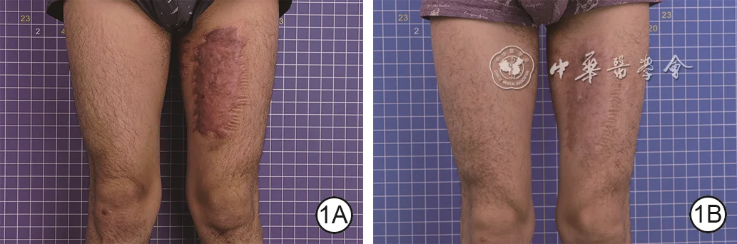
The main goals of treating hypertrophic scars are to reduce redness, firmness, thickening, itching, and pain, and to promote scar maturation. Common methods include silicone gel, silicone sheets, pressure therapy, tension-reduction care, laser treatment, medication injections, and surgical revision. Early intervention is usually easier to control.
 View full-size image
Combined-treatment outcome
A hypertrophic scar of the left thigh was treated with pressure therapy combined with narrow-band intense pulsed light; after nine months, the scar was flatter and less congested.
- 1ABefore treatment, the hypertrophic scar is markedly raised above the skin surface with prominent vascular congestion.
- 1BAfter nine months of treatment, scar flatness has improved and vascular congestion has decreased.
Image source:Zeng Y, Wang C, Lin HY, et al. Clinical efficacy of pressure therapy combined with narrow-band intense pulsed light for hypertrophic scars. Chin J Burns Wounds. 2026;42(4):324–331. DOI: 10.3760/cma.j.cn501225-20251211-00518
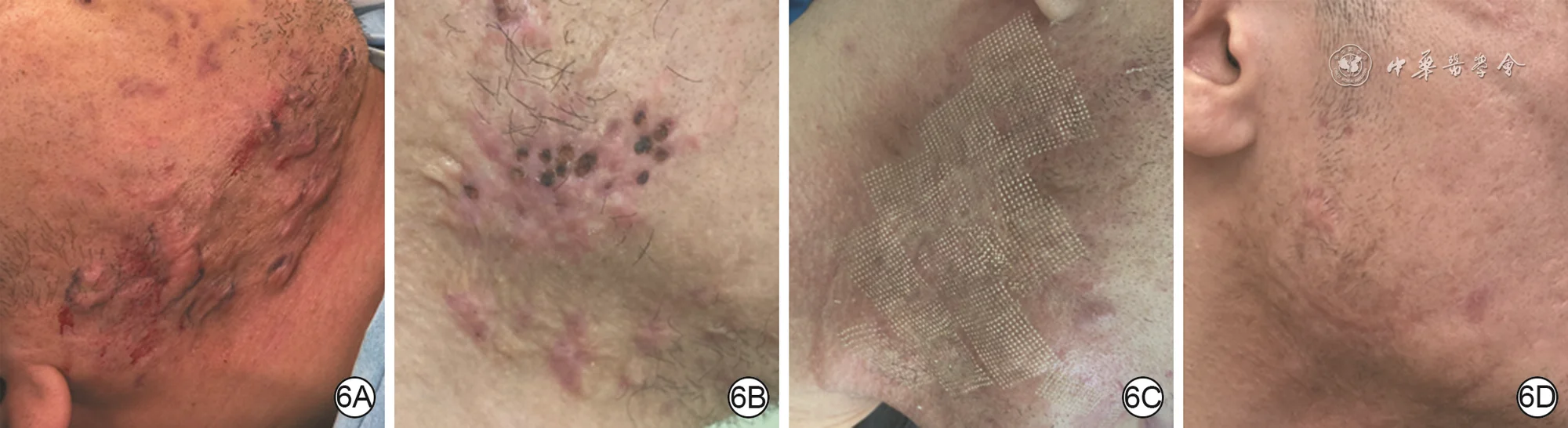
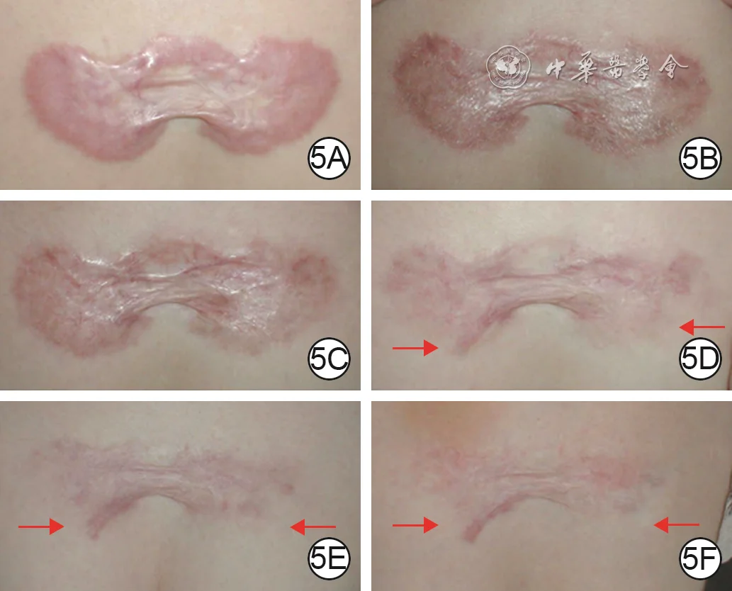
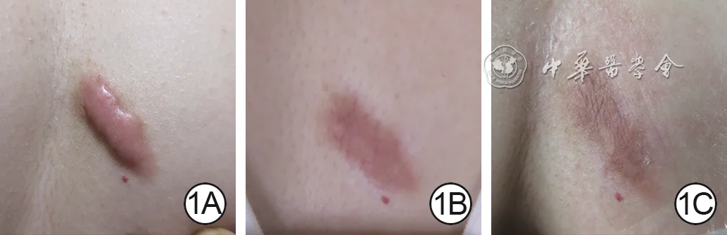
Keloid treatment places greater emphasis on combined therapy and recurrence control. Simple surgical excision has a relatively high risk of recurrence because surgery creates a new wound that may stimulate scar overgrowth again. In clinical practice, depending on the specific situation, surgery combined with radiotherapy, steroid injections, silicone therapy, laser treatment, cryotherapy, or combined medication therapy may be used.
 View full-size image
Staged multimodal remodeling
Multiple scattered right mandibular keloids underwent staged debulking with two punch excisions, followed by fractional carbon dioxide laser to improve punch marks and surface texture; two months after laser treatment, the area was flat and close in color to the surrounding skin.
- 6AAt presentation, multiple scattered keloids are visible in the right mandibular region with an uneven surface.
- 6BOne month after the first punch excision, the keloids are flatter but remain uneven; a second punch excision is then performed.
- 6CTwo months after the second punch excision, the keloids are largely flat but punch marks remain; fractional carbon dioxide laser treatment is then performed.
- 6DTwo months after laser treatment, the scars are essentially removed and the treated area is flat and close to normal skin color.
Image source:Liu W. Application of interventional tissue remodeling strategy in scar prevention and treatment. Chin J Burns Wounds. 2025;41(4):325–332. DOI: 10.3760/cma.j.cn501225-20250102-00004
 View full-size image
Long-term injection remodeling
A plaque-like chest keloid was treated with alternating low-concentration 5-fluorouracil and triamcinolone injections, with concentration and treatment intervals adjusted according to response; fourteen months after treatment cessation, most of the lesion had remodeled toward a normal skin-like appearance.
- 5AAt presentation, a plaque-like keloid shows marked vascular congestion.
- 5BAfter 26 weeks of alternating low-concentration 5-fluorouracil and triamcinolone injections, progression of the keloid is effectively controlled.
- 5CAfter a further eight weeks of injections, the lesion becomes progressively flatter.
- 5DFour months after further adjustment of injection concentration, marked remodeling is visible in the areas indicated by the red arrows.
- 5EAfter nine months of lower concentrations and longer injection intervals, the remodeled areas indicated by the arrows have expanded.
- 5FAt 14 months after treatment cessation, most of the lesion has remodeled toward a normal skin-like appearance; arrows indicate the remodeled extent.
Image source:Liu W, Wu X, Gao Z, et al. Remodelling of keloid tissue into normal-looking skin. J Plast Reconstr Aesthet Surg. 2008;61(12):1553–1554. DOI: 10.1016/j.bjps.2008.08.003
 View full-size image
Treatment follow-up
A post-folliculitis chest keloid treated with dynamically combined pulsed dye laser and triamcinolone injection showed progressive improvement in thickness, vascularity, and color.
- 1ABefore the first treatment, the keloid is markedly elevated above the skin surface with prominent vascular congestion.
- 1BOne month after pulsed dye laser combined with triamcinolone injection, the lesion is thinner and its color has improved.
- 1CAt two months, the keloid is flat with further color improvement, representing a marked response.
Image source:Liu ZN, Zhou YM, Liu RX, et al. Clinical effects of pulsed dye laser dynamically combined with triamcinolone acetonide in the treatment of keloids. Chin J Burns Wounds. 2022;38(9):822–829. DOI: 10.3760/cma.j.cn501225-20220620-00249
 View full-size image
Long-term follow-up
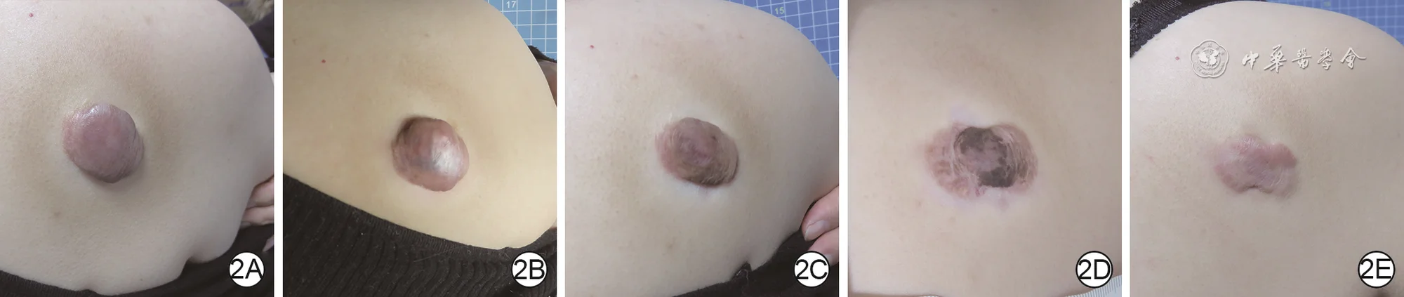
Serial follow-up after triamcinolone injection for a shoulder-back keloid: thickness and color fluctuated early, followed by progressive flattening and fading, with a marked response at ten months.
- 2ABefore the first treatment, the keloid is markedly elevated above the skin surface and dark in color.
- 2BOne month after triamcinolone injection, the lesion is darker and thicker than before treatment.
- 2CAt two months, the keloid is slightly flatter and its color has improved.
- 2DAt seven months, the lesion’s color shows continued improvement.
- 2EAt ten months, the keloid is flat and pale, representing a marked response.
Image source:Liu ZN, Zhou YM, Liu RX, et al. Clinical effects of pulsed dye laser dynamically combined with triamcinolone acetonide in the treatment of keloids. Chin J Burns Wounds. 2022;38(9):822–829. DOI: 10.3760/cma.j.cn501225-20220620-00249
 View full-size image
Preoperative planning
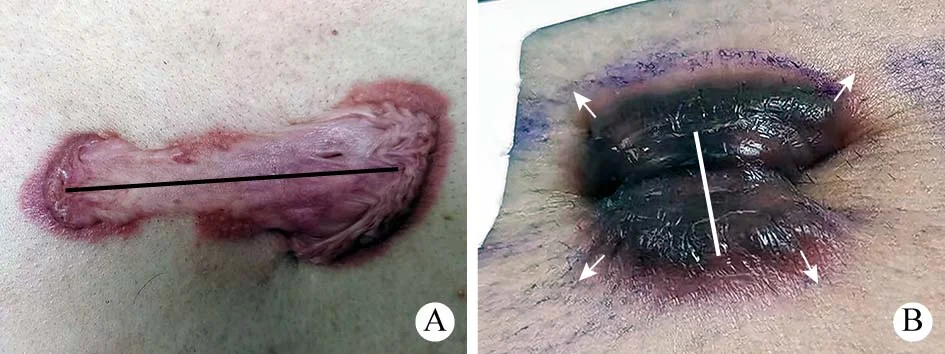
Incision planning for chest-wall keloids incorporates the lesion axis, surrounding skin folds, and local tension direction so that the incision is aligned with regional mechanical forces.
- AFor a manubrial keloid without evident surrounding skin folds, a linear incision is planned along the lesion’s long axis.
- BFor a xiphoid-region keloid, surrounding skin folds indicate the local tension direction (blue arrows); the linear incision is planned parallel to that direction.
Image source:Pan F, Wei BH, Yang W, Guo LL. Clinical outcomes of keloid core excision combined with radiotherapy in treatment of chest keloid. Academic Journal of Chinese PLA Medical School. 2022;43(5):518–523. DOI: 10.3969/j.issn.2095-5227.2022.05.005
 View full-size image
Operative example
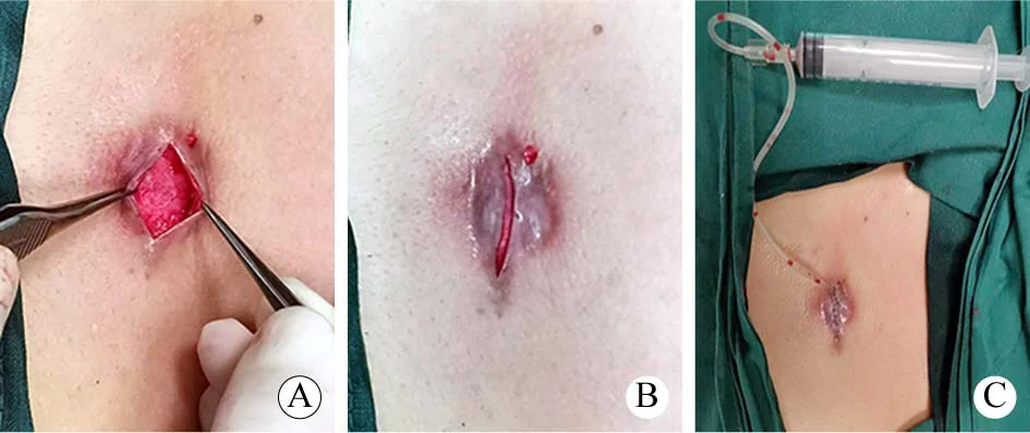
Core excision of a chest-wall keloid with preservation of an approximately 1-mm scar flap, low-tension layered closure, and placement of a subcutaneous negative-pressure drain.
- AThe skin is incised along the long axis of the keloid; the core is dissected and removed while an approximately 1-mm scar flap is preserved.
- BImmediately after core excision, the wound edges can be approximated without obvious tension.
- CThe incision is closed in aligned layers, with a negative-pressure drain placed in the subcutaneous plane.
Image source:Pan F, Wei BH, Yang W, Guo LL. Clinical outcomes of keloid core excision combined with radiotherapy in treatment of chest keloid. Academic Journal of Chinese PLA Medical School. 2022;43(5):518–523. DOI: 10.3969/j.issn.2095-5227.2022.05.005
Pathological Scar Q&A
What should be noted in daily care?
Early wound care affects the final appearance of the scar. Keeping the wound clean, preventing infection, reducing pulling tension, and avoiding repeated friction are important measures to reduce pathological scarring.
After a wound has scabbed over, do not repeatedly pick or scratch it. Do not squeeze acne lesions, especially on areas where keloids commonly occur, such as the chest, jawline, shoulders, and back. Surgical incisions should have dressing changes, suture removal, and follow-up visits according to the doctor’s instructions. For wounds in areas that move, tension-reducing tape or other tension-reducing measures may be used under medical guidance.
After the wound has closed, pay attention to sun protection. Ultraviolet light can worsen pigmentation and make the scar darker. For scars that are prone to overgrowth, silicone gel, silicone sheets, or pressure therapy may be used under medical guidance.
People with a personal or family history of keloids should be cautious about ear piercing, tattoos, body piercings, and non-essential cosmetic skin procedures. When a new skin injury occurs, earlier professional care is recommended.
How can you make an initial self-assessment?
You can make an initial assessment from several aspects:
Whether it extends beyond the original wound A scar confined within the original wound is more consistent with a hypertrophic scar; a scar that extends beyond the original wound and spreads outward is more consistent with a keloid.
Whether it continues to enlarge A scar that gradually stabilizes, softens, and flattens is more like a hypertrophic scar; one that expands over a long period or repeatedly becomes active is more like a keloid.
Where it appears If a firm lump-like overgrowth appears on areas such as the earlobe, front of the chest, shoulders and back, jawline, or upper arm, keloid should be strongly considered.
Whether similar conditions occur in the family If multiple family members are prone to developing keloids, an individual’s risk is increased.
Whether it recurs after excision If it grows back after excision, or even becomes larger than before, the likelihood of a keloid is higher.
These judgments can only serve as a reference. The final assessment still requires a doctor to evaluate the medical history, appearance, palpation findings, course of development, and response to treatment.
When is it advisable to seek medical care early?
If a scar is only mildly red, gradually fading, and not causing obvious discomfort, you may observe it first while providing proper care.
It is advisable to see a plastic surgery department or scar specialist early in the following situations: the scar continues to become thicker, harder, or larger; there is obvious itching, pain, or stinging; it extends beyond the original wound; it is located in a high-risk area such as the earlobe, front of the chest, shoulders, or back; it affects joint movement; it affects the appearance of exposed areas such as the face or neck; a scar rises rapidly after surgery; or a large area of hypertrophic scarring forms after a burn.
The earlier pathological scars are managed, the easier they usually are to control. If they become extensive, very firm, or clearly symptomatic, treatment may take longer and the risk of recurrence may be higher.
References
- New Scarology, edited by Huiyuan Li, Kaihua Lu, and Shuzhong Guo, Fourth Military Medical University Press.
- Monstrey S, et al. Updated scar management practical guidelines: non-invasive and invasive measures. J Plast Reconstr Aesthet Surg. 2014;67(8):1017–1025. PubMed
- Gold MH, et al. Updated international clinical recommendations on scar management: part 2—algorithms for scar prevention and treatment. Dermatol Surg. 2014;40(8):825–831. PubMed
- Zeng Y, Wang C, Lin HY, et al. Clinical efficacy of pressure therapy combined with narrow-band intense pulsed light for hypertrophic scars. Chin J Burns Wounds. 2026;42(4):324–331. DOI: 10.3760/cma.j.cn501225-20251211-00518
- Liu W. Application of interventional tissue remodeling strategy in scar prevention and treatment. Chin J Burns Wounds. 2025;41(4):325–332. DOI: 10.3760/cma.j.cn501225-20250102-00004
- Liu W, Wu X, Gao Z, et al. Remodelling of keloid tissue into normal-looking skin. J Plast Reconstr Aesthet Surg. 2008;61(12):1553–1554. DOI: 10.1016/j.bjps.2008.08.003
- Liu ZN, Zhou YM, Liu RX, et al. Clinical effects of pulsed dye laser dynamically combined with triamcinolone acetonide in the treatment of keloids. Chin J Burns Wounds. 2022;38(9):822–829. DOI: 10.3760/cma.j.cn501225-20220620-00249
- Pan F, Wei BH, Yang W, Guo LL. Clinical outcomes of keloid core excision combined with radiotherapy in treatment of chest keloid. Academic Journal of Chinese PLA Medical School. 2022;43(5):518–523. DOI: 10.3969/j.issn.2095-5227.2022.05.005