There is no single scar treatment that suits everyone. Scars differ in their causes, location, thickness, color, firmness, mobility, symptoms, and risk of recurrence, and the treatment goals are also different.
For example, some people mainly want to improve appearance, some need relief from itching and pain, and some already have functional problems such as joint contracture, eyelid ectropion, restricted mouth opening at the corners of the mouth, or fingers that cannot fully straighten.
When doctors develop a scar treatment plan, they usually first determine the type of scar, then assess the stage the scar is in, and finally choose an appropriate combination of treatments based on the location, individual predisposition, age, treatment risks, and the patient’s expectations.
Step 1: Determine the Scar Type
The doctor will first observe the scar’s appearance. Common types of scars include superficial scars, linear scars, hypertrophic scars, keloids, atrophic scars, depressed scars, contracture scars, webbed scars, bridged scars, and pigmented scars.
Superficial scars mainly present with color changes, with only mild changes in texture and thickness. Linear scars are common after surgical incisions or knife wounds. Hypertrophic scars are usually red, raised, firm, and itchy, but their extent is mostly limited to the original wound. Keloids, by contrast, extend beyond the original area of injury into the surrounding normal skin, and their risk of recurrence is relatively high. Atrophic and depressed scars are common after acne, chickenpox, infection, or trauma. Contracture scars pull on the skin and joints and affect movement and function.
Determining the type is very important. For example, even when scars are similarly raised, hypertrophic scars and keloids require different treatment strategies. Hypertrophic scars can be encouraged to mature through methods such as silicone gel or sheets, pressure therapy, laser treatment, and injections. For keloids, more attention must be paid to the risk of recurrence, and simple excision alone usually carries a higher risk.
Step 2: Determine the Stage of Scar Development
Scars have their own developmental process. Early scars are often reddish, firm, thickened, and itchy, with abundant local blood vessels and active collagen metabolism. This stage is called the active phase or proliferative phase. In the stable phase, the scar color gradually fades, firmness decreases, thickness tends to stabilize, and itching and pain also lessen.
Treatment in the active phase focuses on controlling proliferation and inflammation. Doctors may choose silicone therapy, pressure therapy, laser treatment, intralesional scar injections, tension-reducing care, and other methods to make the scar as soft, flat, and pale as possible.
Treatment in the stable phase is more suitable for contour correction. For example, if wide scars, depressed scars, linear scars, or contracture scars are already relatively stable, the doctor may consider surgical repair, local flaps, skin grafting, fat grafting, subcision, or laser treatment depending on the situation.
Assessing the scar stage can help avoid errors in treatment timing. For example, performing an inappropriate operation too early may stimulate the scar to proliferate again, while delaying treatment of a severe contracture may lead to joint stiffness and reduced function.
Step 3: Assess the Scar Location and Skin Tension
The location of the scar directly affects the choice of treatment. For example, areas such as the anterior chest, shoulders and back, upper arms, jawline, and earlobes are prone to hypertrophic scars and keloids. Areas such as the eyelids, around the mouth, nasal ala, neck, hands, armpits, elbow creases, and knee creases have higher functional and cosmetic requirements.
Skin tension is also a key factor doctors assess. The greater the tension, the more likely a scar is to widen, thicken, and recur. Scars on the chest and shoulders are often subject to pulling forces, so the probability of renewed proliferation after simple excision is relatively high. Scars near joints are repeatedly stressed during movement, so treatment must take into account tension reduction, splints or braces, rehabilitation, and postoperative care.
For facial scars, attention is paid to aesthetic units, skin texture, facial expression movement, and color differences. For hand scars, attention is paid to tendon gliding, sensation, grip strength, and fine motor movements. For neck scars, attention is paid to pulling during head extension, head turning, and swallowing, and so on. Doctors consider both the scar itself and its impact on surrounding structures.
Step 4: Understand the Cause of Scar Formation and Treatment History
Scars from different sources require different treatment approaches. For example, for a surgical incision scar, the incision direction, suture tension, and postoperative care should be considered. For a burn scar, the burn depth, area, healing time, and whether joint regions are involved should be considered. For an acne scar, the type of depression and whether inflammation is still active should be considered. For a traumatic scar, the doctor should assess whether there is a foreign body, infection, tissue loss, or deep adhesion.
Past treatment history is also very important. The doctor will ask whether the patient has had laser treatment, injections, cryotherapy, surgery, radiotherapy, silicone therapy, or pressure therapy; whether there was improvement after treatment; whether the scar recurred; how quickly it recurred; and whether the scar became larger than before treatment.
Step 5: Assess Symptoms and Functional Impact
Scar treatment is not based only on appearance. The doctor will ask about symptoms such as itching, pain, stinging, burning sensations, numbness, tightness, pulling, ulceration, and bleeding. When symptoms are pronounced, this suggests that the scar may still be active, or that local nerve, blood-vessel, inflammatory, and tension-related problems are more prominent.
Functional impact is an important basis for treatment priority. For example, being unable to fully straighten the fingers, difficulty making a fist, inability to raise the shoulder high, limited neck rotation, inability to close the eye completely, difficulty opening the mouth, or impaired walking all suggest that the scar has caused structural pulling. In this situation, the treatment goal should shift from simply improving appearance to restoring function.
Post-burn scars, scars in children, and scars near joints especially require early functional assessment. For example, children are still growing and developing, and scars lack the extensibility of normal skin. If long-term pulling occurs, it may affect development of the limbs and face.
Step 6: Identify People at High Risk of Recurrence
Some patients are more prone to pathological scarring. The doctor will pay attention to skin color, ethnicity, age, family history, previous history of keloids, scar location, and wound-healing status.
People with darker skin, Asians, Black people, and Hispanic people have a relatively higher risk of developing keloids. Adolescents and young adults have active tissue repair and are also more likely to develop hypertrophic scars. If multiple family members have keloids, an individual’s risk also increases.
For high-risk patients, doctors are usually more cautious before surgery, ear piercing, tattooing, body piercing, or cosmetic skin procedures. People who already have a keloid tendency should avoid unnecessary trauma as much as possible. If surgery is necessary, more active preventive measures are often needed after the operation.
Step 7: Define the Treatment Goals
The doctor and patient will clarify the treatment goals together, such as making the scar flatter, softer, and lighter; reducing itching and pain; improving depression and unevenness; narrowing the scar width; releasing pulling and contracture; restoring joint movement; reducing recurrence; and improving the appearance of exposed areas.
Different goals lead to different treatment choices. For example, scars mainly characterized by redness may be prioritized for vascular-related laser treatment; thick, hard, raised scars may be treated with a combination of injections, laser, silicone gel, and pressure therapy; depressed acne scars may be treated with fractional laser, radiofrequency microneedling, subcision, filling, or surgery; when contracture scars affect function, surgical release, skin grafting, flaps, and rehabilitation training may be needed.
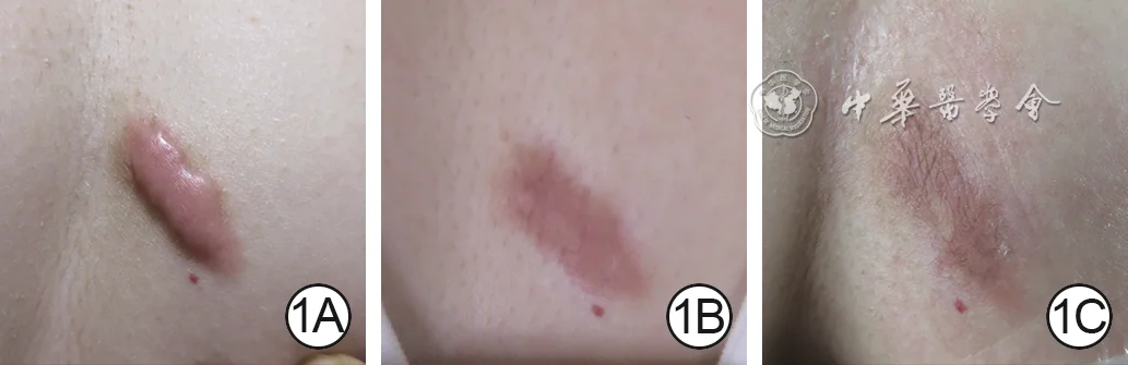 View full-size image
Treatment follow-up
A post-folliculitis chest keloid treated with dynamically combined pulsed dye laser and triamcinolone injection showed progressive improvement in thickness, vascularity, and color.
- 1ABefore the first treatment, the keloid is markedly elevated above the skin surface with prominent vascular congestion.
- 1BOne month after pulsed dye laser combined with triamcinolone injection, the lesion is thinner and its color has improved.
- 1CAt two months, the keloid is flat with further color improvement, representing a marked response.
Image source:Liu ZN, Zhou YM, Liu RX, et al. Clinical effects of pulsed dye laser dynamically combined with triamcinolone acetonide in the treatment of keloids. Chin J Burns Wounds. 2022;38(9):822–829. DOI: 10.3760/cma.j.cn501225-20220620-00249
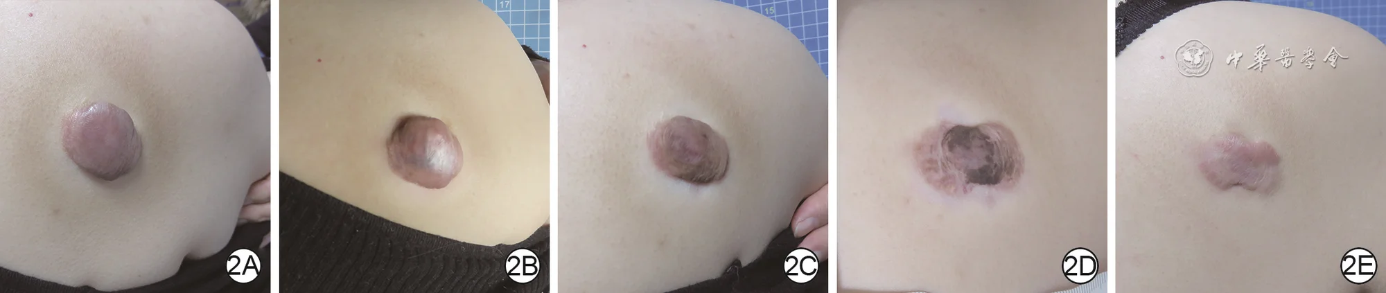 View full-size image
Long-term follow-up
Serial follow-up after triamcinolone injection for a shoulder-back keloid: thickness and color fluctuated early, followed by progressive flattening and fading, with a marked response at ten months.
- 2ABefore the first treatment, the keloid is markedly elevated above the skin surface and dark in color.
- 2BOne month after triamcinolone injection, the lesion is darker and thicker than before treatment.
- 2CAt two months, the keloid is slightly flatter and its color has improved.
- 2DAt seven months, the lesion’s color shows continued improvement.
- 2EAt ten months, the keloid is flat and pale, representing a marked response.
Image source:Liu ZN, Zhou YM, Liu RX, et al. Clinical effects of pulsed dye laser dynamically combined with triamcinolone acetonide in the treatment of keloids. Chin J Burns Wounds. 2022;38(9):822–829. DOI: 10.3760/cma.j.cn501225-20220620-00249
Reasonable expectations are also very important in scar treatment, because mature scars that have already formed are difficult to restore completely to the original normal skin. The goal of medical treatment is usually to improve color, thickness, texture, shape, and function, making the scar less noticeable, more comfortable, and more stable.
Step 8: Choose the Treatment Method
Mild scars, early hypertrophic scars, and scars with less severe symptoms are often treated first with nonsurgical methods. Common approaches include silicone therapy, pressure therapy, laser treatment, intralesional scar injections, cryotherapy, topical medications, moisturizing and barrier repair, sun protection, and tension-reduction care.
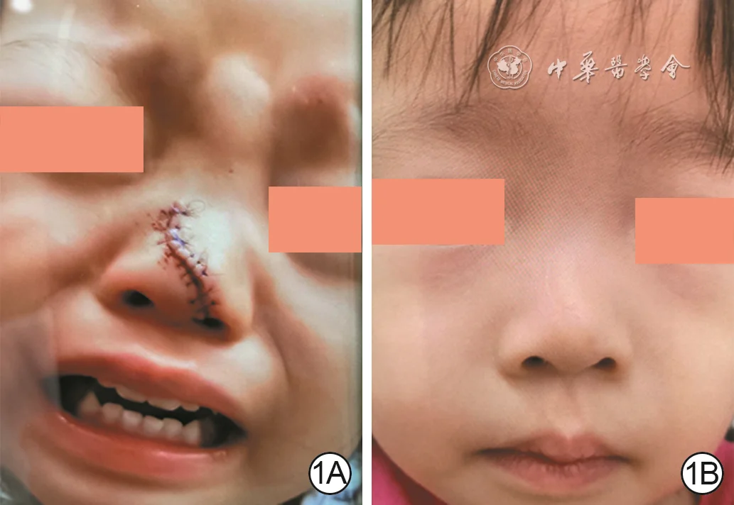 View full-size image
Early-intervention follow-up
Early scar intervention after suturing of a pediatric nasal injury: after three fractional carbon dioxide laser sessions and one needle-knife release of subcutaneous adhesion, the area was flat at 35 weeks and nasal contour and structure were well restored.
- 1AOne week after suturing elsewhere, a linear wound is visible on the lower nasal dorsum; closure is coarse, with mild edge malalignment and uneven height.
- 1BAt 35 weeks after injury, following three fractional carbon dioxide laser sessions and one needle-knife release of subcutaneous adhesion, the wound area is flat and nasal contour and structure are well restored.
Image source:Liu W. Application of interventional tissue remodeling strategy in scar prevention and treatment. Chin J Burns Wounds. 2025;41(4):325–332. DOI: 10.3760/cma.j.cn501225-20250102-00004
Surgical treatment is suitable for scars with more obvious structural problems, such as wide scars, depressed scars, bridged scars, contracture scars, scars affecting facial features or joint function, extensive post-burn scars, and stable scars with limited response to nonsurgical treatment.
Surgical methods include scar excision with meticulous suturing, Z-plasty, W-plasty, local flaps, skin grafting, tissue expansion, regional flaps, and microsurgical free flaps. Which method is chosen depends on the size of the defect, the condition of the surrounding skin, functional needs, and recurrence risk.
Step 9: Develop a Combined Treatment Plan
Scar treatment often requires a combined plan, because a single treatment can address only limited problems, especially for hypertrophic scars, keloids, post-burn scars, and contracture scars.
Common combinations include laser plus injections, surgery plus radiotherapy, surgery plus pressure therapy, surgery plus silicone gel and tension reduction, contracture release plus skin grafting and rehabilitation, and acne-scar treatment combining subcision, laser, and filling.
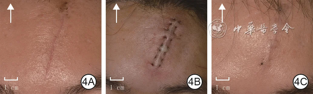 View full-size image
Tension reduction combined with laser treatment
A linear surgical-incision scar of the forehead was treated with horizontal buried intradermal suturing (HBIS) combined with fractional carbon dioxide laser; the scar appearance improved at one month. Arrows indicate the direction of the head.
- 4AThe forehead linear scar before combined treatment; the arrow indicates the direction of the head.
- 4BImmediate appearance after horizontal buried intradermal suturing.
- 4CScar appearance one month after combined fractional carbon dioxide laser treatment.
Image source:Lyu KY, Li YS. Mechanisms of surgical incision scar and technological innovations for scar tension reduction based on the mechano-chemo-biological theory. Chin J Burns Wounds. 2026;42(2):133–142. DOI: 10.3760/cma.j.cn501225-20251013-00424
 View full-size image
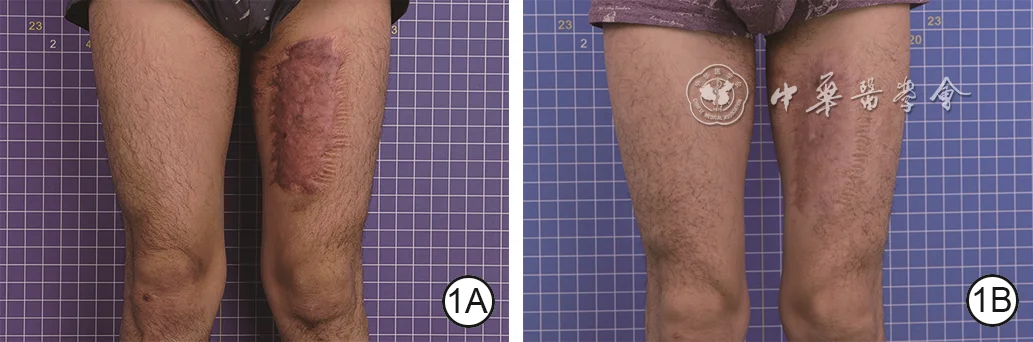
Combined-treatment outcome
A hypertrophic scar of the left thigh was treated with pressure therapy combined with narrow-band intense pulsed light; after nine months, the scar was flatter and less congested.
- 1ABefore treatment, the hypertrophic scar is markedly raised above the skin surface with prominent vascular congestion.
- 1BAfter nine months of treatment, scar flatness has improved and vascular congestion has decreased.
Image source:Zeng Y, Wang C, Lin HY, et al. Clinical efficacy of pressure therapy combined with narrow-band intense pulsed light for hypertrophic scars. Chin J Burns Wounds. 2026;42(4):324–331. DOI: 10.3760/cma.j.cn501225-20251211-00518
 View full-size image
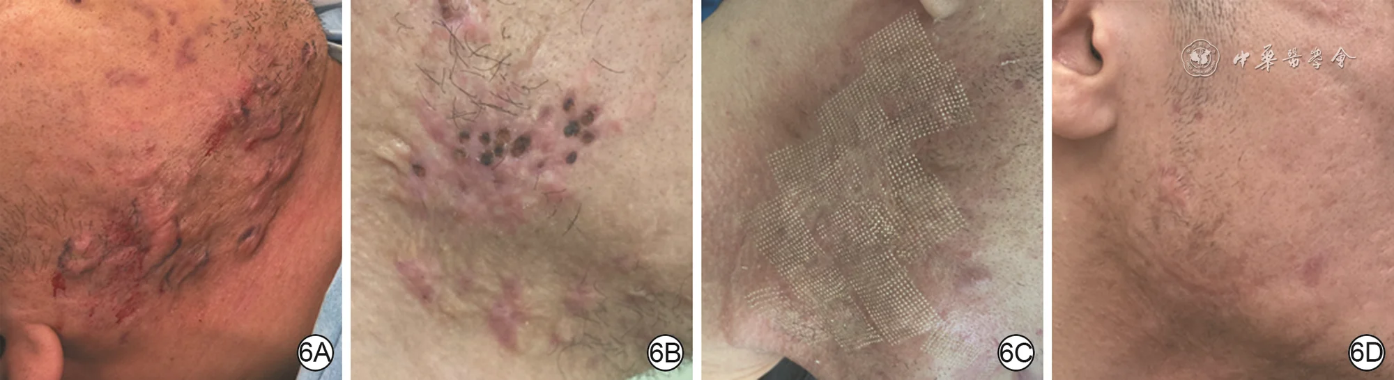
Staged multimodal remodeling
Multiple scattered right mandibular keloids underwent staged debulking with two punch excisions, followed by fractional carbon dioxide laser to improve punch marks and surface texture; two months after laser treatment, the area was flat and close in color to the surrounding skin.
- 6AAt presentation, multiple scattered keloids are visible in the right mandibular region with an uneven surface.
- 6BOne month after the first punch excision, the keloids are flatter but remain uneven; a second punch excision is then performed.
- 6CTwo months after the second punch excision, the keloids are largely flat but punch marks remain; fractional carbon dioxide laser treatment is then performed.
- 6DTwo months after laser treatment, the scars are essentially removed and the treated area is flat and close to normal skin color.
Image source:Liu W. Application of interventional tissue remodeling strategy in scar prevention and treatment. Chin J Burns Wounds. 2025;41(4):325–332. DOI: 10.3760/cma.j.cn501225-20250102-00004
The advantage of combined treatment is that it controls scarring at different stages or mechanisms. For example, injections can suppress overgrowth, laser can improve color and texture, silicone gel and pressure can promote scar maturation, surgery can change the structure, and rehabilitation can restore range of motion.
 View full-size image
Integrated tissue remodeling
An early adult facial laceration was debrided and meticulously resutured, followed after suture removal by ion-beam tissue-remodeling treatment; two months after the second treatment, the wound scar was barely visible.
- 2AAt 3.5 days after the left facial laceration, the closure performed elsewhere appears coarse.
- 2BImmediately after debridement and meticulous resuturing, the wound is flat and well approximated.
- 2CTwo weeks after suture removal, the first ion-beam treatment is administered.
- 2DTwo months after the first ion-beam treatment, the scar is visibly improved and a second treatment is administered.
- 2ETwo months after the second ion-beam treatment, the wound scar is barely visible.
Image source:Liu W. Application of interventional tissue remodeling strategy in scar prevention and treatment. Chin J Burns Wounds. 2025;41(4):325–332. DOI: 10.3760/cma.j.cn501225-20250102-00004
Step 10: Arrange the Treatment Course and Follow-Up
Scar treatment usually requires multiple treatment sessions and long-term follow-up, because laser therapy, injections, pressure therapy, silicone therapy, and similar treatments all need time to take effect. In addition, any new incision after surgery will also go through the process of scar formation and may take several months or even longer to stabilize.
 View full-size image
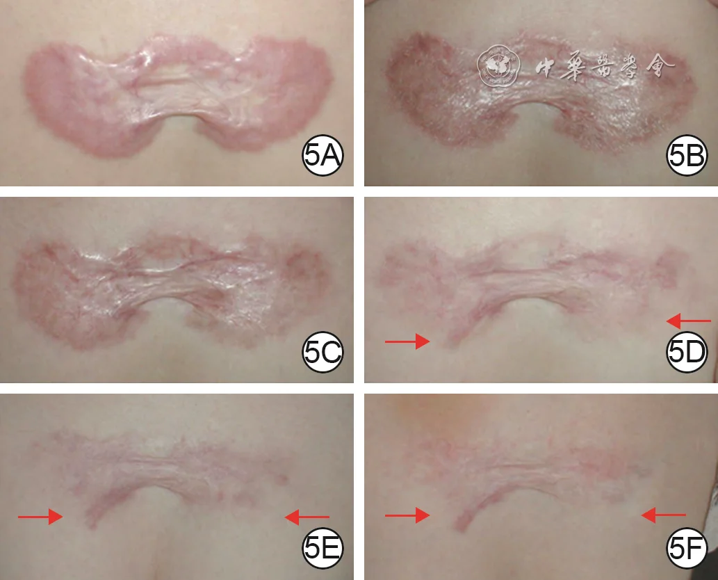
Long-term injection remodeling
A plaque-like chest keloid was treated with alternating low-concentration 5-fluorouracil and triamcinolone injections, with concentration and treatment intervals adjusted according to response; fourteen months after treatment cessation, most of the lesion had remodeled toward a normal skin-like appearance.
- 5AAt presentation, a plaque-like keloid shows marked vascular congestion.
- 5BAfter 26 weeks of alternating low-concentration 5-fluorouracil and triamcinolone injections, progression of the keloid is effectively controlled.
- 5CAfter a further eight weeks of injections, the lesion becomes progressively flatter.
- 5DFour months after further adjustment of injection concentration, marked remodeling is visible in the areas indicated by the red arrows.
- 5EAfter nine months of lower concentrations and longer injection intervals, the remodeled areas indicated by the arrows have expanded.
- 5FAt 14 months after treatment cessation, most of the lesion has remodeled toward a normal skin-like appearance; arrows indicate the remodeled extent.
Image source:Liu W, Wu X, Gao Z, et al. Remodelling of keloid tissue into normal-looking skin. J Plast Reconstr Aesthet Surg. 2008;61(12):1553–1554. DOI: 10.1016/j.bjps.2008.08.003
 View full-size image
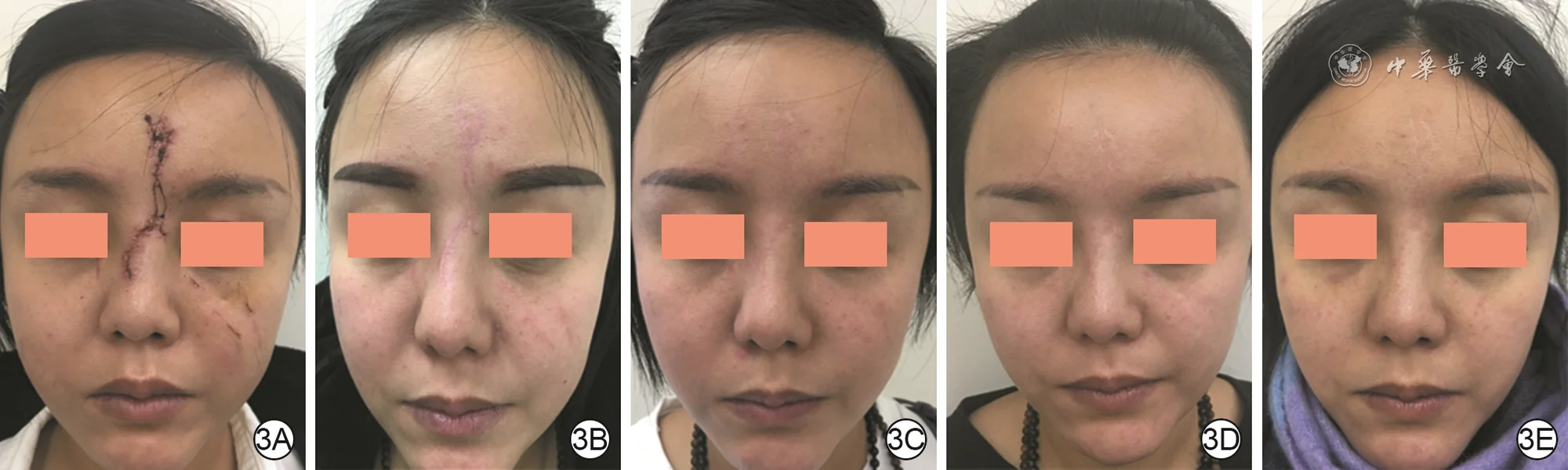
Long-term multimodal intervention
Early adult forehead and nasal wounds underwent long-term multimodal tissue remodeling with oral medication, local injection, ion-beam treatment, and fractional carbon dioxide laser; at 41 weeks after injury, the treated scars were nearly imperceptible.
- 3AAt two weeks after injury, a vertical healing wound is visible across the forehead and nose, with several smaller facial wounds.
- 3BAt 17 weeks, after oral medication, local injection, and two energy-based treatments, wound appearance has improved and a third treatment is administered.
- 3CAt 25 weeks, eight weeks after the third energy-based treatment, the scars are markedly improved and a fourth ion-beam treatment is administered.
- 3DAt 33 weeks, eight weeks after the fourth ion-beam treatment, the forehead and nasal scars show further improvement with fine residual traces; a fifth ion-beam treatment is administered.
- 3EAt 41 weeks, eight weeks after the fifth ion-beam treatment, the scars in the treated area are nearly imperceptible.
Image source:Liu W. Application of interventional tissue remodeling strategy in scar prevention and treatment. Chin J Burns Wounds. 2025;41(4):325–332. DOI: 10.3760/cma.j.cn501225-20250102-00004
During treatment, the doctor will also adjust the plan according to how the scar actually responds. For example, if the scar becomes softer and flatter and symptoms lessen, the original plan may be continued or the interval between treatments may be extended. If the scar continues to thicken, itch, or hurt, injections may be increased, laser settings may be adjusted, or surgery may be considered.
The purpose of follow-up is dynamic management: scars change, and the treatment plan must change with them.
Scar Treatment Q&A
What information does the patient need to provide when the plan is being made?
At the visit, patients can prepare the following information:
the cause of scar formation; the time of injury or surgery; how long the wound took to heal; whether there was infection or ulceration; whether the scar has continued to enlarge; whether there is itching, pain, or tightness; what treatments have been tried before; how effective those treatments were; whether anyone in the family is prone to keloids; what problem the patient most wants to improve; and so on.
Photographs documenting earlier scar changes are also helpful. Patients can provide images from the early and proliferative stages and before and after treatment to show the rate of change. For scars that restrict movement, it is useful to record joint range of motion, difficulties with everyday activities, and pain severity.
References
- New Scarology, edited by Huiyuan Li, Kaihua Lu, and Shuzhong Guo, Fourth Military Medical University Press.
- Monstrey S, et al. Updated scar management practical guidelines: non-invasive and invasive measures. J Plast Reconstr Aesthet Surg. 2014;67(8):1017–1025. PubMed
- Gold MH, et al. Updated international clinical recommendations on scar management: part 2—algorithms for scar prevention and treatment. Dermatol Surg. 2014;40(8):825–831. PubMed
- Lyu KY, Li YS. Mechanisms of surgical incision scar and technological innovations for scar tension reduction based on the mechano-chemo-biological theory. Chin J Burns Wounds. 2026;42(2):133–142. DOI: 10.3760/cma.j.cn501225-20251013-00424
- Zeng Y, Wang C, Lin HY, et al. Clinical efficacy of pressure therapy combined with narrow-band intense pulsed light for hypertrophic scars. Chin J Burns Wounds. 2026;42(4):324–331. DOI: 10.3760/cma.j.cn501225-20251211-00518
- Liu W. Application of interventional tissue remodeling strategy in scar prevention and treatment. Chin J Burns Wounds. 2025;41(4):325–332. DOI: 10.3760/cma.j.cn501225-20250102-00004
- Liu W, Wu X, Gao Z, et al. Remodelling of keloid tissue into normal-looking skin. J Plast Reconstr Aesthet Surg. 2008;61(12):1553–1554. DOI: 10.1016/j.bjps.2008.08.003
- Liu ZN, Zhou YM, Liu RX, et al. Clinical effects of pulsed dye laser dynamically combined with triamcinolone acetonide in the treatment of keloids. Chin J Burns Wounds. 2022;38(9):822–829. DOI: 10.3760/cma.j.cn501225-20220620-00249